. 2019 Jul 2;10(4):e01017-19.
doi: 10.1128/mBio.01017-19.

An Intermolecular π-Stacking Interaction Drives Conformational Changes Necessary to β-Barrel Formation in a Pore-Forming Toxin

Affiliations

An Intermolecular π-Stacking Interaction Drives Conformational Changes Necessary to β-Barrel Formation in a Pore-Forming Toxin

Joshua R Burns et al. mBio. .

Abstract

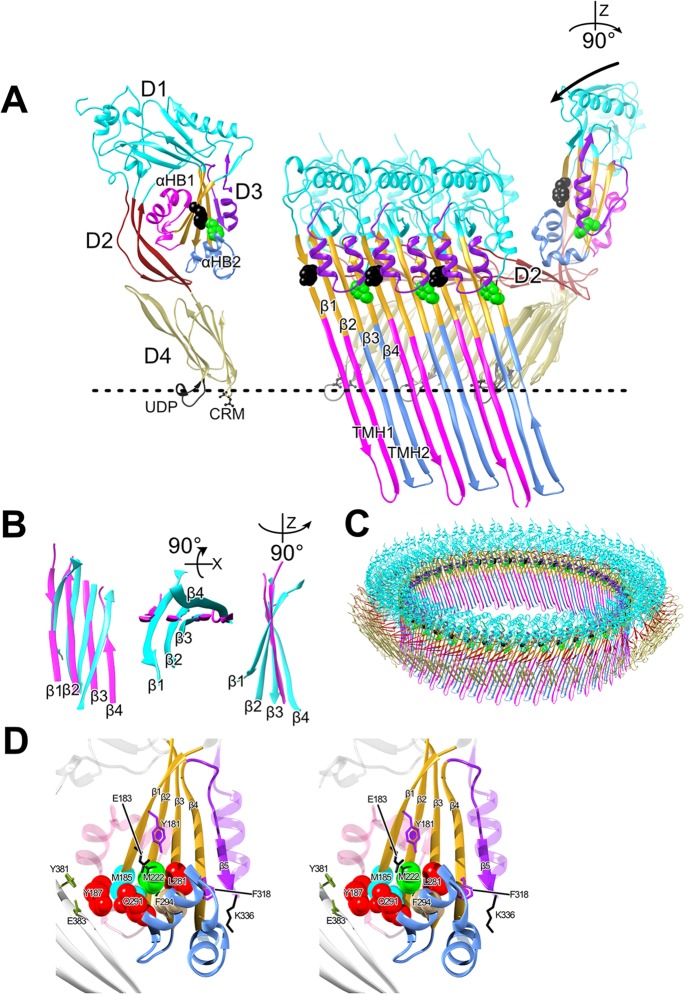
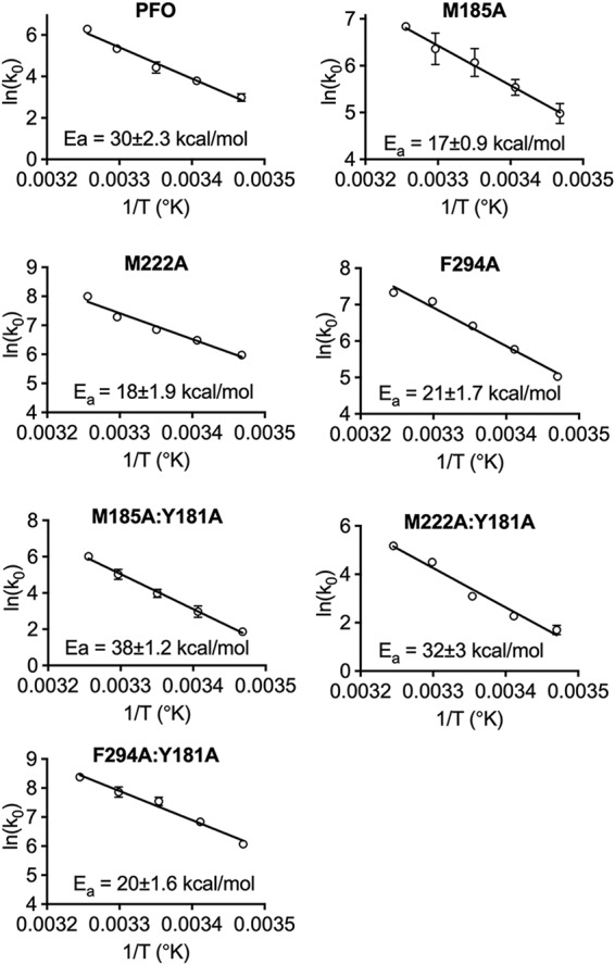
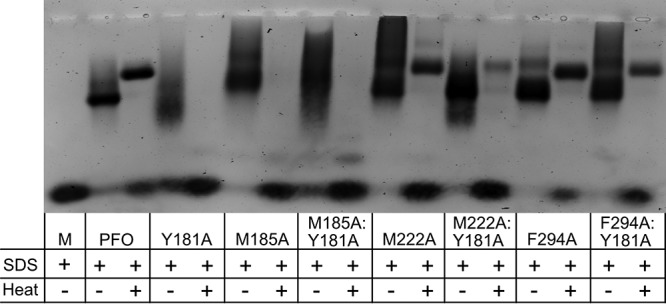
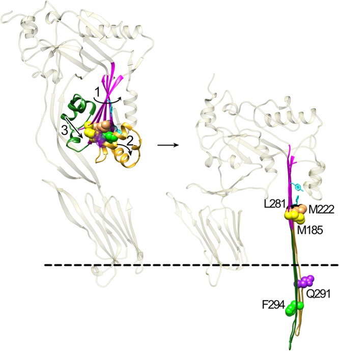
The crystal structures of the soluble monomers of the pore-forming cholesterol-dependent cytolysins (CDCs) contain two α-helical bundles that flank a twisted core β-sheet. This protein fold is the hallmark of the CDCs, as well as of the membrane attack complex/perforin immune defense proteins and the stonefish toxins. To form the β-barrel pore, a core β-sheet is flattened to align the membrane-spanning β-hairpins. Concomitantly with this conformational change, the two α-helical bundles that flank the core β-sheet break their restraining contacts and refold into two membrane-spanning β-hairpins of the β-barrel pore. The studies herein show that in the monomer structure of the archetype CDC perfringolysin O (PFO), a conserved Met-Met-Phe triad simultaneously contributes to maintaining the twist in this core β-sheet, as well as restricting the α-helical-to-β-strand transition necessary to form one of two membrane-spanning β-hairpins. A previously identified intermolecular π-stacking interaction is now shown to disrupt the interactions mediated by this conserved triad. This is required to establish the subsequent intermolecular electrostatic interaction, which has previously been shown to drive the final conformational changes necessary to form the β-barrel pore. Hence, these studies show that the intermolecular π-stacking and electrostatic interactions work in tandem to flatten the core β-sheet and initiate the α-helical-to-β-strand transitions to form the β-barrel pore.IMPORTANCE A unique feature of the CDC/MACPF/SNTX (cholesterol-dependent cytolysin/membrane attack complex perforin/stonefish toxin) superfamily of pore-forming toxins is that the β-strands that comprise the β-barrel pore are derived from a pair of α-helical bundles. These studies reveal the molecular basis by which the formation of intermolecular interactions within the prepore complex drive the disruption of intramolecular interactions within each monomer of the prepore to trigger the α-helical-to-β-strand transition and formation of the β-barrel pore.

Keywords: cholesterol-dependent cytolysin; membrane attack complex; oligomer; perforin; β-barrel.


Figures

FIG 1
FIG 1
PFO structure and mechanism. (A) α-Carbon backbone traces of the crystal structure of the soluble PFO monomer (16) (left panel) and 3 PFO monomers from the pore complex model of PFO (right panel) shown in panel C, which is modeled on the 4.5-Å cryoEM structure of the closely related CDC pneumolysin (20). In the right panel, domain 4 of the soluble monomer structure was aligned with domain 4 of the monomer from the pore to show the ratchet-like lowering via domain 2 of domains 1 and 3 as they move ∼40-Å closer to the membrane (7, 20, 32). The vertical collapse of domain 3 toward the membrane is necessary so that the transmembrane β-hairpins 1 and 2 (TMH1 and -2, magenta and light blue, respectively), which form the β-barrel pore, can cross the membrane bilayer (3, 32) at a 20° tilt (10). Upon prepore-to-pore transition, α-helical bundles 1 and 2 (αHB1 and -2) refold into the membrane-spanning β-hairpins TMH1 and -2 to form the β-barrel pore (2, 3). (B) Overlay of the domain 3 core β-sheets from the soluble PFO monomer structure (16) (cyan) and membrane-inserted PFO monomer from the PLY-based pore model (magenta) showing the flattening of the twist in the β-sheet upon the transition to the pore. Shown left to right are the core β-sheet structures viewed from the interior of the oligomeric complex (a bottom-up view and a side view). (C) PFO pore modeled on the cryoEM-derived 4.5-Å resolution pneumolysin pore (20). (D) Stereo representation of the core β-sheet (gold) and αHBs of domain 3. Shown are the locations of the residues in this study. The dashed line in panels A and C shows the approximate location of the upper surface of the membrane. D1 to D4, domains 1 to 4; CRM, cholesterol recognition/binding motif; UDP; conserved undecapeptide.
FIG 2
FIG 2
Arrhenius activation energies (Eas) of PFO and PFO variants. The initial velocities (k0s) of pore formation for PFO and its derivatives were determined by injecting the toxins directly into a stirred cuvette containing CF liposomes maintained at temperatures (T) from 15 to 35°C. The initial velocities (k0s) of pore formation were used to calculate the Ea. All experiments were performed in triplicate. Standard error bars for all data points are included, although many do not show because they are smaller than the symbols.
FIG 3
FIG 3
Oligomer formation by PFO and PFO variants. Oligomer formation was assessed by incubating PFO and its derivatives with POPC-cholesterol liposomes for 30 min at 37°C. After incubation, sample buffer was added to the proteoliposomes with or without heating them to 95°C for 3 min. Oligomers were resolved on a 1.5% SDS agarose gel. M, PFO monomer without liposomes and heat.
FIG 4
FIG 4
Proposed mechanism by which intermolecular π-stacking and electrostatic interactions drive β-barrel formation. In step 1, the intermolecular π-stacking interaction between Y181 and F318 initially relieves some of the twist in the core β-sheet (magenta), which brings K336 and E183 sufficiently close to form an intermolecular electrostatic interaction. The formation of the electrostatic interaction completes the alignment of β-strands 1 and 4 between two monomers, which flattens the core β-sheet. The progressive flattening of the core β-sheet by the tandem actions of the π-stacking and electrostatic interactions simultaneously disrupts the interactions between F294 and the core β-sheet residues M185, M222, and L281 (step 2) and between αHB1 and domains 1 and 2 (14) (step 3). Both αHB1 and -2, now free of the contacts (or weakened contacts) that maintained their structure, then refold and extend into TMH1 and TMH2, which form the β-barrel pore (2, 10, 11). F294 faces the bilayer, and Q291 faces the pore lumen. M185, M222, and L281 remain lined up in the core β-sheet. F318 is behind L281. The dashed line represents the surface of the bilayer.

References

    1. Sellman BR, Kagan BL, Tweten RK. 1997. Generation of a membrane-bound, oligomerized pre-pore complex is necessary for pore formation by Clostridium septicum alpha toxin. Mol Microbiol 23:551–558. doi:10.1046/j.1365-2958.1997.d01-1876.x. - DOI - PubMed
    1. Shatursky O, Heuck AP, Shepard LA, Rossjohn J, Parker MW, Johnson AE, Tweten RK. 1999. The mechanism of membrane insertion for a cholesterol-dependent cytolysin: a novel paradigm for pore-forming toxins. Cell 99:293–299. doi:10.1016/S0092-8674(00)81660-8. - DOI - PubMed
    1. Shepard LA, Shatursky O, Johnson AE, Tweten RK. 2000. The mechanism of assembly and insertion of the membrane complex of the cholesterol-dependent cytolysin perfringolysin O: formation of a large prepore complex. Biochemistry 39:10284–10293. doi:10.1021/bi000436r. - DOI - PubMed
    1. Walker B, Braha O, Cheley S, Bayley H. 1995. An intermediate in the assembly of a pore-forming protein trapped with a genetically-engineered switch. Chem Biol 2:99–105. doi:10.1016/1074-5521(95)90282-1. - DOI - PubMed
    1. Miller CJ, Elliot JL, Collier RL. 1999. Anthrax protective antigen: prepore-to-pore conversion. Biochemistry 38:10432–10441. doi:10.1021/bi990792d. - DOI - PubMed

Publication types

MeSH terms

Substances