

Differences in the Binding Affinity of an HIV-1 V2 Apex-Specific Antibody for the SIVsmm/mac Envelope Glycoprotein Uncouple Antibody-Dependent Cellular Cytotoxicity from Neutralization
- PMID: 31266872
- PMCID: PMC6606807
- DOI: 10.1128/mBio.01255-19
Differences in the Binding Affinity of an HIV-1 V2 Apex-Specific Antibody for the SIVsmm/mac Envelope Glycoprotein Uncouple Antibody-Dependent Cellular Cytotoxicity from Neutralization
Abstract
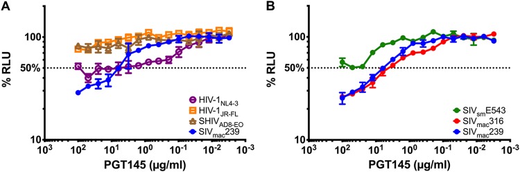
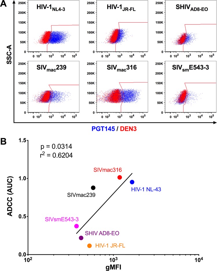
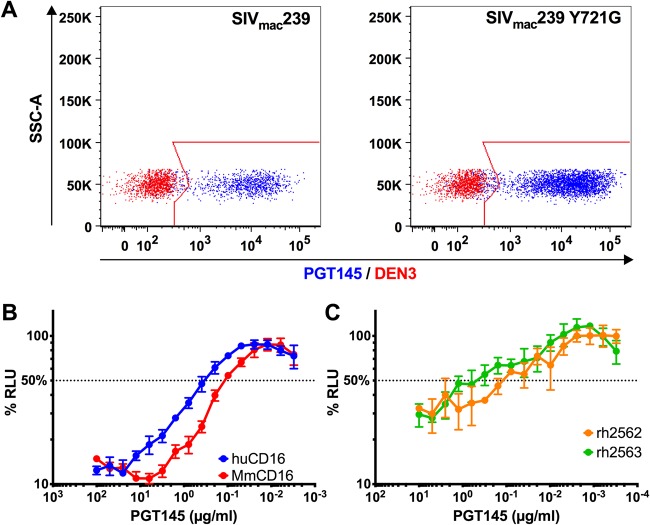
As a consequence of their independent evolutionary origins in apes and Old World monkeys, human immunodeficiency virus type 1 (HIV-1) and simian immunodeficiency viruses of the SIVsmm/mac lineage express phylogenetically and antigenically distinct envelope glycoproteins. Thus, HIV-1 Env-specific antibodies do not typically cross-react with the Env proteins of SIVsmm/mac isolates. Here we show that PGT145, a broadly neutralizing antibody to a quaternary epitope at the V2 apex of HIV-1 Env, directs the lysis of SIVsmm/mac-infected cells by antibody-dependent cellular cytotoxicity (ADCC) but does not neutralize SIVsmm/mac infectivity. Amino acid substitutions in the V2 loop of SIVmac239 corresponding to the epitope for PGT145 in HIV-1 Env modulate sensitivity to this antibody. Whereas a substitution in a conserved N-linked glycosylation site (N171Q) eliminates sensitivity to ADCC, a lysine-to-serine substitution in this region (K180S) increases ADCC and renders the virus susceptible to neutralization. These differences in function correlate with an increase in the affinity of PGT145 binding to Env on the surface of virus-infected cells and to soluble Env trimers. To our knowledge, this represents the first instance of an HIV-1 Env-specific antibody that cross-reacts with SIVsmm/mac Env and illustrates how differences in antibody binding affinity for Env can differentiate sensitivity to ADCC from neutralization.IMPORTANCE Here we show that PGT145, a potent broadly neutralizing antibody to HIV-1, directs the lysis of SIV-infected cells by antibody-dependent cellular cytotoxicity but does not neutralize SIV infectivity. This represents the first instance of cross-reactivity of an HIV-1 Env-specific antibody with SIVsmm/mac Env and reveals that antibody binding affinity can differentiate sensitivity to ADCC from neutralization.
Keywords: ADCC; antibody function; human immunodeficiency virus; neutralizing antibodies; simian immunodeficiency virus.
Copyright © 2019 von Bredow et al.
Figures
Similar articles
-
Antibody-dependent cellular cytotoxicity, infected cell binding and neutralization by antibodies to the SIV envelope glycoprotein.PLoS Pathog. 2023 May 30;19(5):e1011407. doi: 10.1371/journal.ppat.1011407. eCollection 2023 May. PLoS Pathog. 2023. PMID: 37253062 Free PMC article.
-
Potent antibody-dependent cellular cytotoxicity of a V2-specific antibody is not sufficient for protection of macaques against SIV challenge.PLoS Pathog. 2024 Jan 22;20(1):e1011819. doi: 10.1371/journal.ppat.1011819. eCollection 2024 Jan. PLoS Pathog. 2024. PMID: 38252675 Free PMC article.
-
Predominant envelope variable loop 2-specific and gp120-specific antibody-dependent cellular cytotoxicity antibody responses in acutely SIV-infected African green monkeys.Retrovirology. 2018 Mar 9;15(1):24. doi: 10.1186/s12977-018-0406-5. Retrovirology. 2018. PMID: 29523166 Free PMC article.
-
Comparison of Antibody-Dependent Cell-Mediated Cytotoxicity and Virus Neutralization by HIV-1 Env-Specific Monoclonal Antibodies.J Virol. 2016 Jun 10;90(13):6127-6139. doi: 10.1128/JVI.00347-16. Print 2016 Jul 1. J Virol. 2016. PMID: 27122574 Free PMC article.
-
Nonneutralizing functional antibodies: a new "old" paradigm for HIV vaccines.Clin Vaccine Immunol. 2014 Aug;21(8):1023-36. doi: 10.1128/CVI.00230-14. Epub 2014 Jun 11. Clin Vaccine Immunol. 2014. PMID: 24920599 Free PMC article. Review.
Cited by
-
Mycobacterium tuberculosis disease associates with higher HIV-1-specific antibody responses.iScience. 2023 Apr 10;26(5):106631. doi: 10.1016/j.isci.2023.106631. eCollection 2023 May 19. iScience. 2023. PMID: 37168567 Free PMC article.
-
Antibody-dependent cellular cytotoxicity, infected cell binding and neutralization by antibodies to the SIV envelope glycoprotein.PLoS Pathog. 2023 May 30;19(5):e1011407. doi: 10.1371/journal.ppat.1011407. eCollection 2023 May. PLoS Pathog. 2023. PMID: 37253062 Free PMC article.
-
V2-Specific Antibodies in HIV-1 Vaccine Research and Natural Infection: Controllers or Surrogate Markers.Vaccines (Basel). 2019 Aug 6;7(3):82. doi: 10.3390/vaccines7030082. Vaccines (Basel). 2019. PMID: 31390725 Free PMC article. Review.
-
Molecular insights into antibody-mediated protection against the prototypic simian immunodeficiency virus.Nat Commun. 2022 Sep 6;13(1):5236. doi: 10.1038/s41467-022-32783-2. Nat Commun. 2022. PMID: 36068229 Free PMC article.
-
An Automated Fluorescence-Based Method to Isolate Bone Marrow-Derived Plasma Cells from Rhesus Macaques Using SIVmac239 SOSIP.664.Mol Ther Methods Clin Dev. 2020 Aug 5;18:781-790. doi: 10.1016/j.omtm.2020.08.004. eCollection 2020 Sep 11. Mol Ther Methods Clin Dev. 2020. PMID: 32953929 Free PMC article.
References
-
- Keele BF, Jones JH, Terio KA, Estes JD, Rudicell RS, Wilson ML, Li Y, Learn GH, Beasley TM, Schumacher-Stankey J, Wroblewski E, Mosser A, Raphael J, Kamenya S, Lonsdorf EV, Travis DA, Mlengeya T, Kinsel MJ, Else JG, Silvestri G, Goodall J, Sharp PM, Shaw GM, Pusey AE, Hahn BH. 2009. Increased mortality and AIDS-like immunopathology in wild chimpanzees infected with SIVcpz. Nature 460:515–519. doi:10.1038/nature08200. - DOI - PMC - PubMed
-
- Apetrei C, Kaur A, Lerche NW, Metzger M, Pandrea I, Hardcastle J, Falkenstein S, Bohm R, Koehler J, Traina-Dorge V, Williams T, Staprans S, Plauche G, Veazey RS, McClure H, Lackner AA, Gormus B, Robertson DL, Marx PA. 2005. Molecular epidemiology of simian immunodeficiency virus SIVsm in U.S. primate centers unravels the origin of SIVmac and SIVstm. J Virol 79:8991–9005. doi:10.1128/JVI.79.14.8991-9005.2005. - DOI - PMC - PubMed
Publication types
MeSH terms
Substances
Grants and funding
LinkOut - more resources
Full Text Sources
Other Literature Sources
Research Materials
Miscellaneous
