. 2019 Jul 2;10(1):2928.
doi: 10.1038/s41467-019-10975-7.

Transient drug-tolerance and permanent drug-resistance rely on the trehalose-catalytic shift in Mycobacterium tuberculosis

Affiliations

Transient drug-tolerance and permanent drug-resistance rely on the trehalose-catalytic shift in Mycobacterium tuberculosis

Jae Jin Lee et al. Nat Commun. .

Abstract

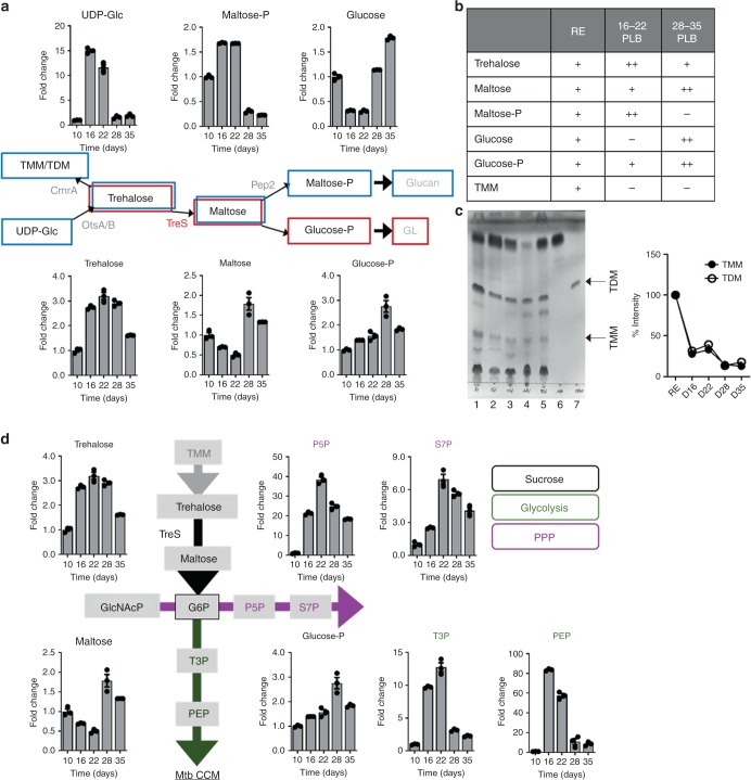
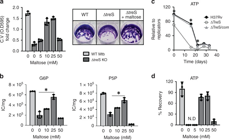
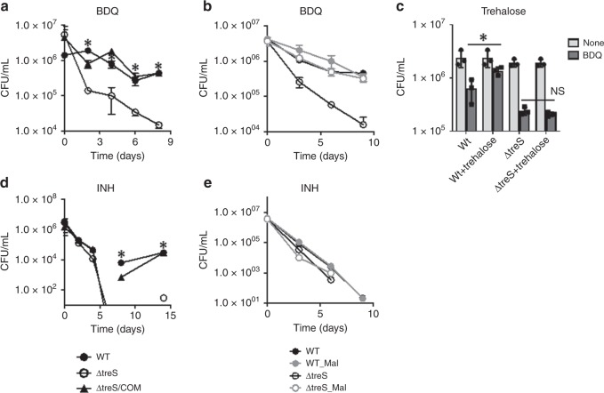
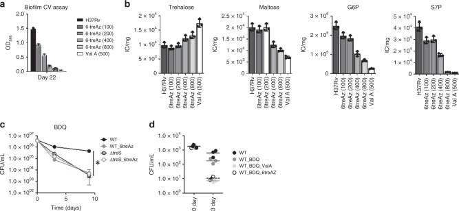
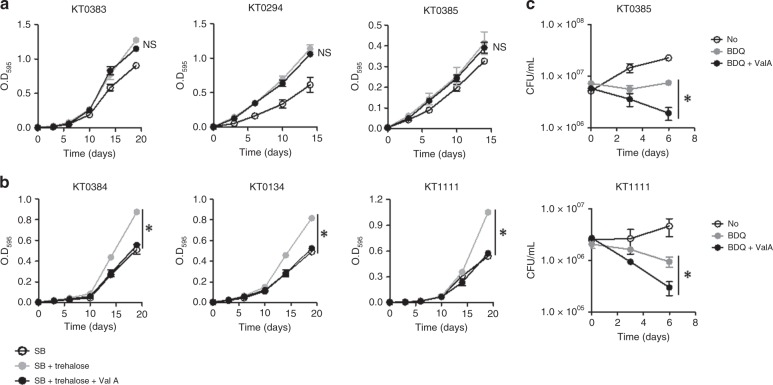
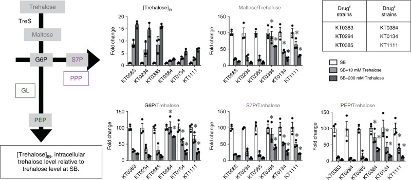
Stochastic formation of Mycobacterium tuberculosis (Mtb) persisters achieves a high level of antibiotic-tolerance and serves as a source of multidrug-resistant (MDR) mutations. As conventional treatment is not effective against infections by persisters and MDR-Mtb, novel therapeutics are needed. Several approaches were proposed to kill persisters by altering their metabolism, obviating the need to target active processes. Here, we adapted a biofilm culture to model Mtb persister-like bacilli (PLB) and demonstrated that PLB underwent trehalose metabolism remodeling. PLB use trehalose as an internal carbon to biosynthesize central carbon metabolism intermediates instead of cell surface glycolipids, thus maintaining levels of ATP and antioxidants. Similar changes were identified in Mtb following antibiotic-treatment, and MDR-Mtb as mechanisms to circumvent antibiotic effects. This suggests that trehalose metabolism is associated not only with transient drug-tolerance but also permanent drug-resistance, and serves as a source of adjunctive therapeutic options, potentiating antibiotic efficacy by interfering with adaptive strategies.


Conflict of interest statement

The authors declare no competing interests.

Figures

Fig. 1
Fig. 1
Mtb PLB-specific remodeling of trehalose metabolism, glycolysis, and the pentose phosphate pathway. a, d Intermediates of trehalose metabolism were as described in Supplementary Fig. 4. Intrabacterial pool sizes of Mtb intermediates in trehalose metabolism, glycolysis, and the pentose phosphate pathway in PLB harvested from mycobacterial biofilm culture at days 10, 16, 22, 28, and 35. Total bar heights indicate the intrabacterial pool sizes relative to those of PLB at day 10. Blue-boxed metabolites denote the carbon flux of the replicating state and red-boxed metabolites denote the carbon flux of PLB at day 28. UDP-Glc, uracil diphosphate-glucose; maltose-P, maltose phosphate; P5P, pentose 5-phosphates; S7P, sedoheptulose 7-phosphates; G6P, glucose 6-phosphates; T3P, triose 3-phosphates (dihydroxyacetone phosphate and glyceraldehyde phosphate); PEP, phosphoenolpyruvate. b Major changes in time-course PLB metabolomics (one plus, levels in replicating state; two pluses, increased; one minus, decreased). c Analysis of the extractable trehalose monomycolate (TMM), trehalose dimycolate (TDM), and free mycolic acids from PLB at the same time points as metabolome extraction. Equal amounts of total cellular lipids were run in the solvent system (chloroform:methanol:H2O, 90:10:1; v/v/v) (left panel) and TMM/TDM intensity (density) was monitored by ImageJ software (right panel). The labeled band shown in the TLC corresponds to the Rf of a TMM, TDM, or mycolic acid standard. Lane 1, replicating; Lane 2, day 16 PLB; Lane 3, day 22 PLB; Lane 4, day 28 PLB; Lane 5, day 35 PLB; Lane 6, standard mycolic acid; Lane 7, standard TDM. d Black font metabolites are members of sucrose metabolism, green font metabolites are members of glycolysis, and magenta font metabolites are members of the pentose phosphate pathway (PPP). CCM, central carbon metabolism. All values are the average of two independent biological duplicates ± s.e.m. Source data are provided as a Source Data file
Fig. 2
Fig. 2
Metabolic consequences of TreS deficiency on PLB metabolism. a Time-course kinetics of pool sizes of glycolysis (green font/line), the pentose phosphate pathway (magenta font/line), and sucrose metabolism (black font/line) intermediates of WT Mtb, ΔtreS, and ΔtreS/com of mycobacterial biofilm culture. P5P, pentose 5-phosphates; S7P, sedoheptulose 7-phosphates; G6P, glucose 6-phosphates; T3P, triose 3-phosphates (dihydroxyacetone phosphate and glyceraldehyde phosphate); PEP, phosphoenolpyruvate. b Time course of extractable TMM/TDM from WT Mtb, ΔtreS, and ΔtreS/com harvested during the replicating state (I), day 22 (II), and day 28 (III) of mycobacterial biofilm culture. TMM/TDM extraction and TLC development and visualization were conducted as described in Fig. 1c. All values are the average of two independent biological duplicates ±s.e.m. Source data are provided as a Source Data file
Fig. 3
Fig. 3
Maltose-chemical rescue of ΔtreS PLB formation. a CV staining OD595 values of 28-day-old ΔtreS PLB fraction generated within mycobacterial biofilm culture in the presence of 0–50 mM maltose (left panel), and CV staining patterns of WT Mtb, ΔtreS, and ΔtreS treated with 20 mM maltose (right panel). b The effects of supplementing various concentrations of maltose on the ΔtreS PLB metabolome. Total bar heights indicate intrabacterial pool sizes compared with ΔtreS with no maltose supplement or WT Mtb. c Time course of intrabacterial ATP levels of WT Mtb (H37Rv), ΔtreS, and ΔtreS/com while incubating within mycobacterial biofilm culture. d The effect of supplementing ΔtreS PLB fraction with 20 mM maltose on intrabacterial ATP levels at day 28. All values are the average of biological triplicates ± s.e.m. *P < 0.001 by Student’s unpaired t-test. G6P, glucose 6-phosphates; P5P, pentose 5-phosphates. Source data are provided as a Source Data file
Fig. 4
Fig. 4
The trehalose-catalytic shift is used to circumvent BDQ and INH effects. a CFU (colony-forming unit) viability of WT Mtb, ΔtreS, and ΔtreS/com following treatment with 10× MIC-equivalent BDQ. The effect of supplementing with b 20 mM maltose and c trehalose on WT Mtb ΔtreS, and ΔtreS/com CFU viability following treatment with 10× MIC-equivalent BDQ. d CFU viability of WT Mtb, ΔtreS, and ΔtreS/com following treatment with 10× MIC-equivalent INH. e The effect of supplementation of 20 mM maltose on CFU viability of WT Mtb, ΔtreS, and ΔtreS/com with co-treatment of 10× MIC-equivalent INH. All values are the average of experimental triplicates ±s.e.m. and representative of at least two independent experiments. *P < 0.01. NS, not significant by Student’s unpaired t-test
Fig. 5
Fig. 5
Anti-PLB formation activity of 6-azido-6-α,α′-trehalose (6-treAz). a The effects of various concentrations (100–800 µM) of 6-treAz treatment on PLB formation were monitored by CV staining assay of PLB harvested at day 28 of mycobacterial biofilm culture. Treatment with 500 µM validamycin A was included as a positive control. b Metabolic impact of various concentrations of 6-treAz on PLB metabolism. The metabolome of PLB was harvested at day 28 PLB within mycobacterial biofilm culture. Intrabacterial pool sizes of PLB intermediates in trehalose metabolism (trehalose and maltose) and GL (G6P) and PPP (S7P) following treatment with various concentrations of 6-treAz or 500 µM Val A. c CFU-based viability of WT Mtb or ΔtreS in the presence or absence of 200 µM 6-treAz following co-treatment with 10× MIC equivalent of BDQ. d The effect of 6-treAz and/or Val A on CFU-based viability of WT Mtb inside human PBMC macrophages following treatment with 10× MIC-equivalent BDQ. All values are the average of biological triplicates ±s.e.m. *P < 0.001 by ANOVA with Bonferroni post-test correction. S7P, sedoheptulose 7-phosphates; G6P, glucose 6-phosphates. Source data are provided as a Source Data file
Fig. 6
Fig. 6
XDR- and TDR-TB clinical isolates use the trehalose-catalytic shift for survival. a The growth of drug-sensitive TB isolates (KT0294, KT0383, and KT0385) in m7H9 media containing 10 mM sodium butyrate (SB) was enhanced by supplementing with 20 mM trehalose, but not restored by treating with 200 µM validamycin A (Val A). b The growth of drug-resistant isolates (XDR-TB, KT0134, and KT0384; TDR-TB, KT1111) in m7H9 media containing 10 mM SB was enhanced by supplementing with 20 mM trehalose, and completely restored to trehalose-untreated levels following treatment with 200 µM Val A. c Chemical inhibition of TreS as an adjunctive therapeutic strategy to boost BDQ-mediated antimicrobial effect on drug-sensitive and drug-resistant clinical isolates. All values are the average of biological triplicates ±s.e.m. *P < 0.01; NS, not significant by ANOVA between SB + trehalose and SB + trehalose + Val A (a) and between BDQ and BDQ + Val A (b) with Bonferroni post-test correction
Fig. 7
Fig. 7
The effect of supplementing trehalose on TB clinical isolate metabolism. Supplementation with 10 or 20 mM trehalose on the central metabolic pathways of drug-sensitive, DrugS, (KT0294, KT0383, and KT0385), and drug-resistant, DrugR, XDR- (KT0134 and KT0384), and TDR- (KT1111) TB clinical isolates was monitored by metabolomics. Left panel, trehalose metabolism, glycolysis (GL), and the pentose phosphate pathway (PPP) intermediates were analyzed. Right panel, [Trehalose]IB showed intrabacterial trehalose detected within each TB clinical isolate strain following exposure to 0, 10, or 20 mM trehalose supplementation for 24 h. Metabolite fold change describes the percent conversion rate using intake trehalose under each condition, including 0, 10, and 20 mM trehalose supplementation. The conversion rate of each metabolite from trehalose under SB (sodium butyrate with 0 mM trehalose) was set to 100%. All values are the average of biological triplicates ±s.e.m. *P < 0.01 by Student’s unpaired t-test (between drug-resistant TB isolate metabolites and the average of the corresponding metabolite of drug-sensitive TB isolates; KT0294, KT0384, and KT0385). Data shown represent the combined results of duplicates of three replicates. Source data are provided as a Source Data file

References

    1. WHO. Global tuberculosis report (2017).
    1. Jacobson KR. Tuberculosis. Ann. Intern. Med. 2017;166:ITC17–ITC32. doi: 10.7326/AITC201702070. - DOI - PubMed
    1. Pai M, et al. Tuberculosis. Nat. Rev. Dis. Primers. 2016;2:16076. doi: 10.1038/nrdp.2016.76. - DOI - PubMed
    1. Gomez JE, McKinney JD. M. tuberculosis persistence, latency, and drug tolerance. Tuberculosis. 2004;84:29–44. doi: 10.1016/j.tube.2003.08.003. - DOI - PubMed
    1. Nathan C. Taming tuberculosis: a challenge for science and society. Cell Host Microbe. 2009;5:220–224. doi: 10.1016/j.chom.2009.02.004. - DOI - PubMed

Publication types

MeSH terms