. 2019 Aug;36(4):241-257.
doi: 10.1007/s10719-019-09876-0. Epub 2019 Jul 2.

"Stuck on sugars - how carbohydrates regulate cell adhesion, recognition, and signaling"

Affiliations

"Stuck on sugars - how carbohydrates regulate cell adhesion, recognition, and signaling"

Richard D Cummings. Glycoconj J. 2019 Aug.

Abstract

We have explored the fundamental biological processes by which complex carbohydrates expressed on cellular glycoproteins and glycolipids and in secretions of cells promote cell adhesion and signaling. We have also explored processes by which animal pathogens, such as viruses, bacteria, and parasites adhere to glycans of animal cells and initiate disease. Glycans important in cell signaling and adhesion, such as key O-glycans, are essential for proper animal development and cellular differentiation, but they are also involved in many pathogenic processes, including inflammation, tumorigenesis and metastasis, and microbial and parasitic pathogenesis. The overall hypothesis guiding these studies is that glycoconjugates are recognized and bound by a growing class of proteins called glycan-binding proteins (GBPs or lectins) expressed by all types of cells. There is an incredible variety and diversity of GBPs in animal cells involved in binding N- and O-glycans, glycosphingolipids, and proteoglycan/glycosaminoglycans. We have specifically studied such molecular determinants recognized by selectins, galectins, and many other C-type lectins, involved in leukocyte recruitment to sites of inflammation in human tissues, lymphocyte trafficking, adhesion of human viruses to human cells, structure and immunogenicity of glycoproteins on the surfaces of human parasites. We have also explored the molecular basis of glycoconjugate biosynthesis by exploring the enzymes and molecular chaperones required for correct protein glycosylation. From these studies opportunities for translational biology have arisen, involving production of function-blocking antibodies, anti-glycan specific antibodies, and synthetic glycoconjugates, e.g. glycosulfopeptides, that specifically are recognized by GBPs. This invited short review is based in part on my presentation for the IGO Award 2019 given by the International Glycoconjugate Organization in Milan.

Keywords: Antibodies; Cosmc; Glycobiology; Glycomics; Glycoproteins; Immunity; Inflammation; Parasites; Selectins; T-synthase.


Figures

Figure 1.
Figure 1.
Complex carbohydrates on glycoproteins and glycolipids within and on cells and in cellular secretions can be bound by glycan-binding proteins (GBPs) and antibodies, as well as cross-recognized by microbes and viruses and their glycans and GBPs, e.g. adhesins and hemagglutinins. Through these direct (and indirect) interactions, glycans can signal cells, regulate cell adhesion, and participate in a wide range of developmental, immunological, hematological, and cellular/tissue pathways. Alteration or disruption of glycosylation pathways or GBP expression, through acquired and heritable disorders, or drug treatments, and also in tumor cells, typically leads to pathological outcomes.
Figure 2.
Figure 2.
The biosynthesis of mucin-type O-glycans is initiated post-endoplasmic reticulum and in the Golgi apparatus by the addition of GalNAc by a family of ppGalNAcT enzymes, then subsequently galactose is added by the T-synthase, which requires the molecular chaperone Cosmc in the ER for the correct folding and activity of the enzyme. Subsequent modifications of the Thomsen or T antigen (also called the Thomsen-Friedenreich or TF antigen) occurs by additional enzymatic reactions to generate an incredible diversity of O-glycans, including those with the core 2 O-glycan and SLex determinant recognized by P-selectin and other selectins. At the bottom of the figure is a depiction of how P-selectin binds residues within the SLex determinant along with sulfated tyrosine residues and peptide determinants at the extreme N-terminus of PSGL-1.
Figure 3.
Figure 3.
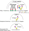
Several examples are shown in which multiple determinants, including glycan, peptide, and tyrosine sulfate, within glycoprotein ligands contribute to high affinity binding of recognition molecules, such as glycan-binding proteins and chemokines.
Figure 4.
Figure 4.
Examples of antigenic glycan determinants expressed in glycoproteins, Asn- or N-glycans of parasitic helminths, including Schistosoma mansoni, and many trematodes and nematodes. The determinants are in colored boxes and their common names are shown, e.g. LDN, FLDNF, etc.
Figure 5.
Figure 5.
The technology of shotgun glycomics requires the release of glycans from biological samples of glycoproteins and glycolipids by enzymatic or chemical strategies. The released glycans, which represent the glycome of the source material, can be fluorescently tagged with a bifunctional linker, and then separated by multi-dimensional chromatography and quantified. The separated glycans are preserved in a tagged-glycan library or TGL, from which they can be covalently or non-covalently printed to generate shotgun glycan microarrays or other presentations. The interrogation of the shotgun glycan microarrays with a GBP, lectin, toxin, or virus, for example, can lead to the identification of a novel set of glycans carrying the determinants needed for recognition. The structural characterization of the glycans retrieved from the TGL can be performed by MS and other technologies. Depicted is the concept of shotgun glycomics applied to influenza virus, where the starting material may be the human lung[213].
Figure 6.
Figure 6.
A depiction of our working model as to how Cosmc functions in the endoplasmic reticulum as a molecular chaperone to assist in folding of newly synthesized T-synthase. Human Cosmc (shown in green), is present in the ER as a disulfide-bonded oligomer, where it can interact with the nascent polypeptide of human T-synthase (shown in red) as it is synthesized. This interaction is reversible and prevents the undesirable association and oligomerization of the T-synthase and inactive forms. The interaction occurs through the Cosmc Binding Region within T-synthase (CBRT), which is accessible in the immature protein but inaccessible once the T-synthase has completed folding and is active. The absence of Cosmc leads to these inactive forms that are proteolyzed eventually in the ER and in the 26S proteasome in the cytoplasm. When Cosmc functions normally, the T-synthase becomes an active dimer that moves to the Golgi apparatus, where it functions to generate normal O-glycans that have core 1 as a precursor with galactose linked to N-acetylgalactosamine. In the absence of Cosmc cells generate the Tn and Sialyl Tn antigen lacking galactose.

References

    1. Cummings RD, Schnaar RL, Esko JD, Drickamer K, Taylor ME: Principles of Glycan Recognition In: Varki A, Cummings RD, Esko JD, Stanley P, Hart GW, Aebi M, Darvill AG, Kinoshita T, Packer NH, Prestegard JH, Schnaar RL, Seeberger PH (eds.) Essentials of Glycobiology. pp. 373–385. Cold Spring Harbor; (NY: ) (2017)
    1. Thompson AJ, de Vries RP, Paulson JC: Virus recognition of glycan receptors. Curr Opin Virol 34, 117–129 (2019) - PMC - PubMed
    1. Kaltner H, Toegel S, Caballero GG, Manning JC, Ledeen RW, Gabius HJ: Galectins: their network and roles in immunity/tumor growth control. Histochem Cell Biol 147(2), 239–256 (2017) - PubMed
    1. Johannes L, Jacob R, Leffler H: Galectins at a glance. J Cell Sci 131(9) (2018). - PubMed
    1. Mendez-Huergo SP, Blidner AG, Rabinovich GA: Galectins: emerging regulatory checkpoints linking tumor immunity and angiogenesis. Curr Opin Immunol 45, 8–15 (2017) - PubMed

Publication types

LinkOut - more resources