

Basal ganglia calcifications (Fahr's syndrome): related conditions and clinical features
- PMID: 31267306
- PMCID: PMC6817747
- DOI: 10.1007/s10072-019-03998-x
Basal ganglia calcifications (Fahr's syndrome): related conditions and clinical features
Erratum in
-
Correction to: Basal ganglia calcifications (Fahr's syndrome): related conditions and clinical features.Neurol Sci. 2019 Nov;40(11):2265. doi: 10.1007/s10072-019-04037-5. Neurol Sci. 2019. PMID: 31444730 Free PMC article.
Abstract
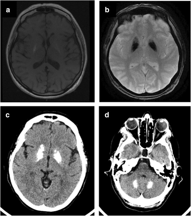
Basal ganglia calcifications could be incidental findings up to 20% of asymptomatic patients undergoing CT or MRI scan. The presence of neuropsychiatric symptoms associated with bilateral basal ganglia calcifications (which could occur in other peculiar brain structures, such as dentate nuclei) identifies a clinical picture defined as Fahr's Disease. This denomination mainly refers to idiopathic forms in which no metabolic or other underlying causes are identified. Recently, mutations in four different genes (SLC20A2, PDGFRB, PDGFB, and XPR1) were identified, together with novel mutations in the Myogenic Regulating Glycosylase gene, causing the occurrence of movement disorders, cognitive decline, and psychiatric symptoms. On the other hand, secondary forms, also identified as Fahr's syndrome, have been associated with different conditions: endocrine abnormalities of PTH, such as hypoparathyroidism, other genetically determined conditions, brain infections, or toxic exposure. The underlying pathophysiology seems to be related to an abnormal calcium/phosphorus homeostasis and transportation and alteration of the blood-brain barrier.
Keywords: Basal ganglia; Brain calcinosis; Fahr’s syndrome; Hypoparathyroidism.
Conflict of interest statement
The author declares that she has no conflict of interest.
Figures
References
-
- Manyam BV. What is and what is not ‘Fahr’s disease’. Parkinsonism Relat Disord. 2005;11:73–80. - PubMed
-
- Yamada M, Asano T, Okamoto K, Hayashi Y, Kanematsu M, Hoshi H, Akaiwa Y, Shimohata T, Nishizawa M, Inuzuka T, Hozumi I. High frequency of calcification in basal ganglia on brain computed tomography images in Japanese older adults. Geriatr Gerontol Int. 2013;13(3):706–710. - PubMed
-
- Simoni M, Pantoni L, Pracucci G, Palmertz B, Guo X, Gustafson D, Skoog I. Prevalence of CT-detected cerebral abnormalities in an elderly Swedish population sample. Acta Neurol Scand. 2008;118(4):260–267. - PubMed
-
- Fahr T. Idiopathische verkalkung der hirngefässe. Zentrabl Allg Pathol. 1930;50:129–133.
Publication types
MeSH terms
Supplementary concepts
LinkOut - more resources
Full Text Sources
Medical
Miscellaneous
