Comment
. 2019 Sep 1;137(9):1029-1036.
doi: 10.1001/jamaophthalmol.2019.2442.

A Quantitative Severity Scale for Retinopathy of Prematurity Using Deep Learning to Monitor Disease Regression After Treatment

Affiliations
Comment

A Quantitative Severity Scale for Retinopathy of Prematurity Using Deep Learning to Monitor Disease Regression After Treatment

Kishan Gupta et al. JAMA Ophthalmol. .

Abstract

Importance: Retinopathy of prematurity (ROP) is a leading cause of childhood blindness worldwide, but treatment failure and disease recurrence are important causes of adverse outcomes in patients with treatment-requiring ROP (TR-ROP).

Objectives: To apply an automated ROP vascular severity score obtained using a deep learning algorithm and to assess its utility for objectively monitoring ROP regression after treatment.

Design, setting, and participants: This retrospective cohort study used data from the Imaging and Informatics in ROP consortium, which comprises 9 tertiary referral centers in North America that screen high volumes of at-risk infants for ROP. Images of 5255 clinical eye examinations from 871 infants performed between July 2011 and December 2016 were assessed for eligibility in the present study. The disease course was assessed with time across the numerous examinations for patients with TR-ROP. Infants born prematurely meeting screening criteria for ROP who developed TR-ROP and who had images captured within 4 weeks before and after treatment as well as at the time of treatment were included.

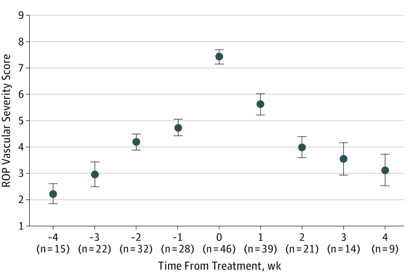
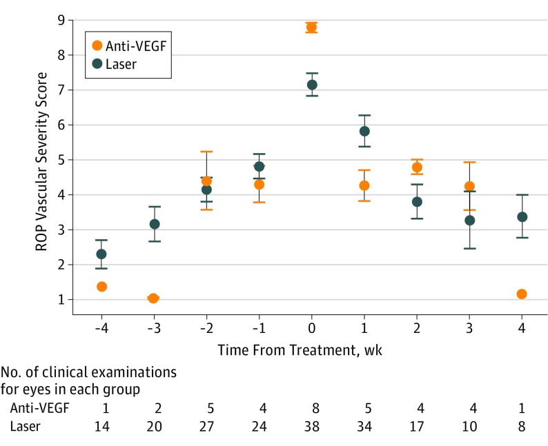
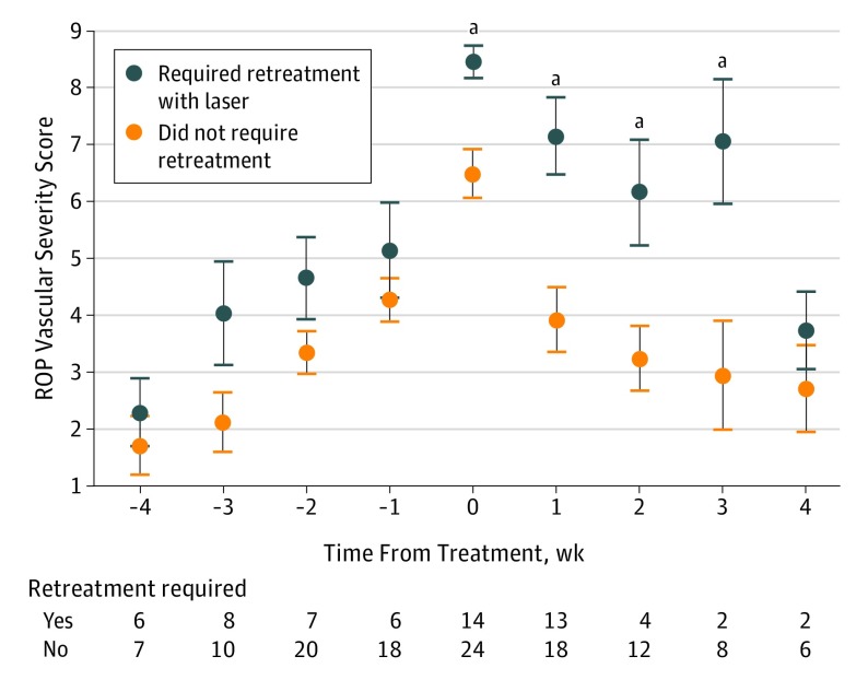
Main outcomes and measures: The primary outcome was mean (SD) ROP vascular severity score before, at time of, and after treatment. A deep learning classifier was used to assign a continuous ROP vascular severity score, which ranged from 1 (normal) to 9 (most severe), at each examination. A secondary outcome was the difference in ROP vascular severity score among eyes treated with laser or the vascular endothelial growth factor antagonist bevacizumab. Differences between groups for both outcomes were assessed using unpaired 2-tailed t tests with Bonferroni correction.

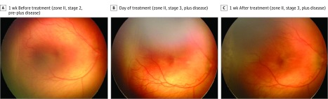
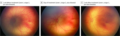
Results: Of 5255 examined eyes, 91 developed TR-ROP, of which 46 eyes met the inclusion criteria based on the available images. The mean (SD) birth weight of those patients was 653 (185) g, with a mean (SD) gestational age of 24.9 (1.3) weeks. The mean (SD) ROP vascular severity scores significantly increased 2 weeks prior to treatment (4.19 [1.75]), peaked at treatment (7.43 [1.89]), and decreased for at least 2 weeks after treatment (4.00 [1.88]) (all P < .001). Eyes requiring retreatment with laser had higher ROP vascular severity scores at the time of initial treatment compared with eyes receiving a single treatment (P < .001).

Conclusions and relevance: This quantitative ROP vascular severity score appears to consistently reflect clinical disease progression and posttreatment regression in eyes with TR-ROP. These study results may have implications for the monitoring of patients with ROP for treatment failure and disease recurrence and for determining the appropriate level of disease severity for primary treatment in eyes with aggressive disease.


Conflict of interest statement

Conflict of Interest Disclosures: Dr Campbell reported receiving grants from Genentech and personal fees from Allergan outside the submitted work. Dr Chan reported receiving personal fees from Alcon, Allergan, Visunex Medical Systems, Beyeonics, and Genentech outside the submitted work. Dr Dy reported receiving grants from the National Science Foundation. Dr Ioannidis reported receiving grants from National Science Foundation and from the National Institutes of Health during the conduct of the study. Dr Kalpathy-Cramer reported receiving grants from the National Eye Institute and the National Science Foundation during the conduct of the study and receiving personal fees from INFOTECHSoft outside the submitted work. Dr Chiang reported receiving grants from the National Institutes of Health, the National Science Foundation, and Genentech; receiving nonfinancial support from Clarity Medical Systems; receiving personal fees from Novartis during the conduct of the study; and being an equity owner in Initeleretina outside the submitted work. Drs Campbell and Brown, Ms Ostmo, and Drs Chan, Dy, Erdogmus, Ioannidis, Kalpathy-Cramer, and Chiang have a patent application pending on the described technology.

Figures

Figure 1.
Figure 1.. Serial Wide-Angle Retinal Images and Corresponding Clinical Diagnosis From Deep Learning in an Infant Treated With Laser
The infant was born at 25 weeks 3 days gestational age and developed treatment-requiring disease. The automated retinopathy of prematurity severity scores are 5.91 (A), 8.01 (B), and 6.99 (C).
Figure 2.
Figure 2.. Serial Wide-Angle Retinal Images and Corresponding Clinical Diagnosis From Deep Learning in an Infant Treated With Intravitreal Bevacizumab
The infant was born at 24 weeks 5 days gestational age and developed treatment-requiring disease. The automated retinopathy of prematurity severity scores are 6.21 (A), 8.94 (B), and 6.09 (C).
Figure 3.
Figure 3.. Quantitative Retinopathy of Prematurity (ROP) Vascular Severity Score Progression and Regression in a Cohort of Infants Who Developed Treatment-Requiring Disease
Mean scores are displayed relative to time of treatment (which is time 0). Number of clinical examinations (n) evaluated at each time is displayed. Error bars indicate 95% CIs.
Figure 4.
Figure 4.. Quantitative Retinopathy of Prematurity (ROP) Vascular Severity Score Distribution in a Cohort of Infants Who Were Treated With Laser Photocoagulation vs Antivascular Endothelial Growth Factor (Anti-VEGF) Agents
Mean scores are displayed 4 weeks prior to, before, and after treatment. Error bars indicate 95% CIs.
Figure 5.
Figure 5.. Quantitative Retinopathy of Prematurity (ROP) Vascular Severity Score Distribution in a Cohort of Infants With Treatment-Requiring Disease Who Received Laser Photocoagulation and Who Required Retreatment With Laser vs Those Who Required No Retreatment
Mean scores are displayed 4 weeks before and after treatment, and the number of clinical examinations is shown at each time. At treatment and 1, 2, and 3 weeks after treatment, retreated eyes had significantly higher ROP vascular severity scores than eyes that never required retreatment. Error bars indicate 95% CIs. aP < .05.

Comment on

References

    1. Cryotherapy for Retinopathy of Prematurity Cooperative Group Multicenter trial of cryotherapy for retinopathy of prematurity: preliminary results. Arch Ophthalmol. 1988;106(4):471-479. doi:10.1001/archopht.1988.01060130517027 - DOI - PubMed
    1. Gilbert C. Retinopathy of prematurity: a global perspective of the epidemics, population of babies at risk and implications for control. Early Hum Dev. 2008;84(2):77-82. doi:10.1016/j.earlhumdev.2007.11.009 - DOI - PubMed
    1. The Committee for the Classification of Retinopathy of Prematurity An international classification of retinopathy of prematurity. Arch Ophthalmol. 1984;102(8):1130-1134. doi:10.1001/archopht.1984.01040030908011 - DOI - PubMed
    1. International Committee for the Classification of Retinopathy of Prematurity The international classification of retinopathy of prematurity revisited. Arch Ophthalmol. 2005;123(7):991-999. doi:10.1001/archopht.123.7.991 - DOI - PubMed
    1. Early Treatment For Retinopathy Of Prematurity Cooperative Group Revised indications for the treatment of retinopathy of prematurity: results of the early treatment for retinopathy of prematurity randomized trial. Arch Ophthalmol. 2003;121(12):1684-1694. doi:10.1001/archopht.121.12.1684 - DOI - PubMed