

Association of Midlife Hearing Impairment With Late-Life Temporal Lobe Volume Loss
- PMID: 31268512
- PMCID: PMC6613307
- DOI: 10.1001/jamaoto.2019.1610
Association of Midlife Hearing Impairment With Late-Life Temporal Lobe Volume Loss
Abstract
Importance: Hearing impairment (HI) in midlife (45-65 years of age) may be associated with longitudinal neurodegeneration of temporal lobe structures, a biomarker of early Alzheimer disease.
Objective: To evaluate the association of midlife HI with brain volume trajectories in later life (≥65 years of age).
Design, setting, and participants: This prospective cohort study used data from the Baltimore Longitudinal Study of Aging to evaluate hearing from November 5, 1990, to October 3, 1994, and late-life volume change from July 10, 2008, to January 29, 2015, using magnetic resonance imaging (MRI) (mean follow-up time, 19.3 years). Data analysis was performed from September 22, 2017, to August 27, 2018. A total of 194 community-dwelling older adults who had midlife measures of peripheral hearing at a mean age of 54.5 years and late-life volume change of up to 6 years between the first and most recent MRI assessment were studied. Excluded were those with baseline cognitive impairment, stroke, head injuries, Parkinson disease, and bipolar disorder.
Exposures: Hearing as measured with pure tone audiometry in each ear from November 5, 1990, to October 3, 1994, and late-life temporal lobe volume change measured by MRI.
Main outcomes and measures: Linear mixed-effects models with random intercepts were used to examine the association of midlife hearing (pure tone average of 0.5-4 kHz tones in the better ear and each ear separately) with longitudinal late-life MRI-based measures of temporal lobe structures (hippocampus, entorhinal cortex, parahippocampal gyrus, and superior, middle, and inferior temporal gyri) in the left and right hemispheres, in addition to global and lobar regions, adjusting for baseline demographic characteristics (age, sex, subsequent cognitive impairment status, and educational level) and intracranial volume.
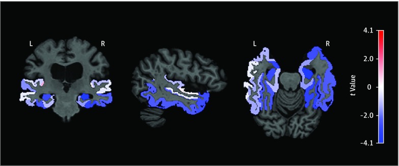
Results: A total of 194 patients (mean [SD] age at hearing assessment, 54.5 [10.0] years; 106 [54.6%] female; 169 [87.1%] white) participated in the study. After Bonferroni correction, poorer midlife hearing in the better ear was associated with steeper late-life volumetric declines in the right temporal gray matter (β = -0.113; 95% CI, -0.182 to -0.044), right hippocampus (β = -0.008; 95% CI, -0.012 to -0.004), and left entorhinal cortex (β = -0.009; 95% CI, -0.015 to -0.003). Poorer midlife hearing in the right ear was associated with steeper late-life volumetric declines in the right temporal gray matter (β = -0.136; 95% CI, -0.197 to -0.075), right hippocampus (β = -0.008; 95% CI, -0.012 to -0.004), and left entorhinal cortex (β = -0.009; 95% CI, -0.015 to -0.003), whereas there were no associations between poorer midlife hearing in the left ear with late-life volume loss.
Conclusions and relevance: The findings suggest that midlife HI is a risk factor for temporal lobe volume loss. Poorer midlife hearing, particularly in the right ear, was associated with declines in hippocampus and entorhinal cortex.
Conflict of interest statement
Figures
References
-
- Villemagne VL, Burnham S, Bourgeat P, et al. ; Australian Imaging Biomarkers and Lifestyle (AIBL) Research Group . Amyloid β deposition, neurodegeneration, and cognitive decline in sporadic Alzheimer’s disease: a prospective cohort study. Lancet Neurol. 2013;12(4):357-367. doi:10.1016/S1474-4422(13)70044-9 - DOI - PubMed
-
- Reiman EM, Quiroz YT, Fleisher AS, et al. . Brain imaging and fluid biomarker analysis in young adults at genetic risk for autosomal dominant Alzheimer’s disease in the presenilin 1 E280A kindred: a case-control study. Lancet Neurol. 2012;11(12):1048-1056. doi:10.1016/S1474-4422(12)70228-4 - DOI - PMC - PubMed
-
- Jack CR Jr, Lowe VJ, Weigand SD, et al. ; Alzheimer’s Disease Neuroimaging Initiative . Serial PIB and MRI in normal, mild cognitive impairment and Alzheimer’s disease: implications for sequence of pathological events in Alzheimer’s disease. Brain. 2009;132(Pt 5):1355-1365. doi:10.1093/brain/awp062 - DOI - PMC - PubMed
