Review
. 2019 Oct;1454(1):14-30.
doi: 10.1111/nyas.14138. Epub 2019 Jul 3.

Vagal innervation of the stomach reassessed: brain-gut connectome uses smart terminals

Affiliations
Review

Vagal innervation of the stomach reassessed: brain-gut connectome uses smart terminals

Terry L Powley et al. Ann N Y Acad Sci. 2019 Oct.

Abstract

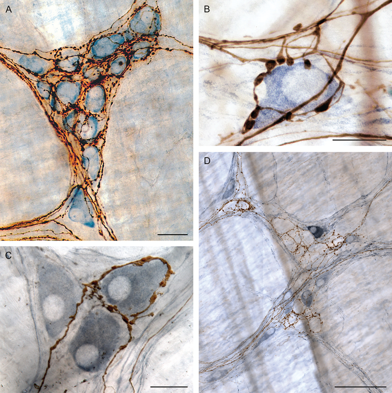
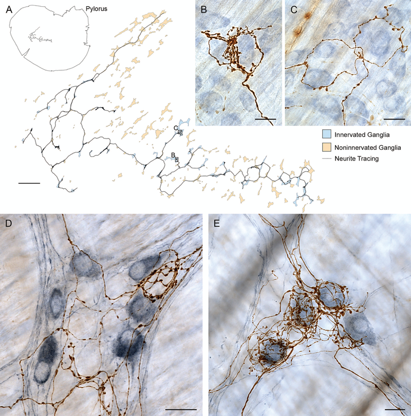
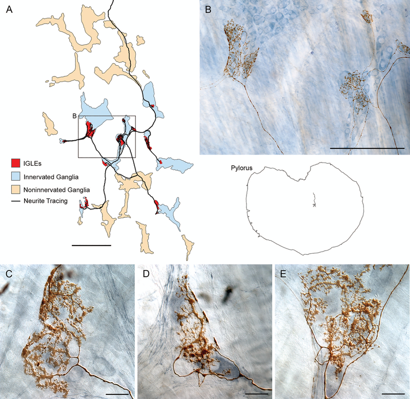
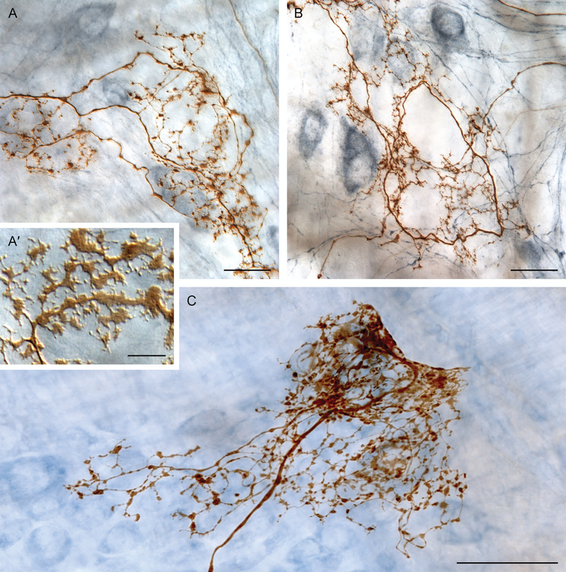
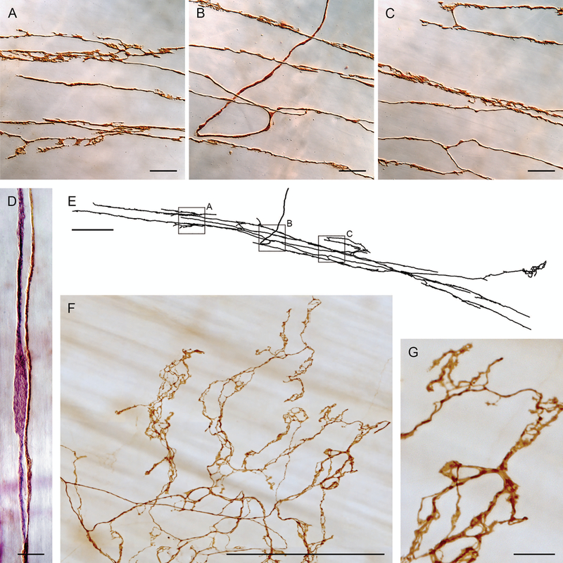
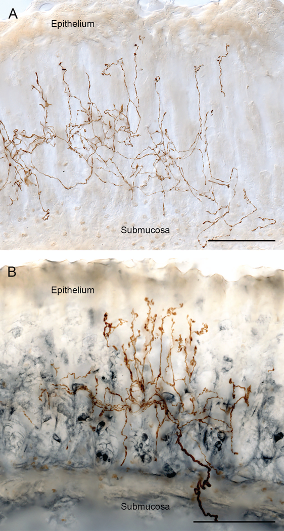
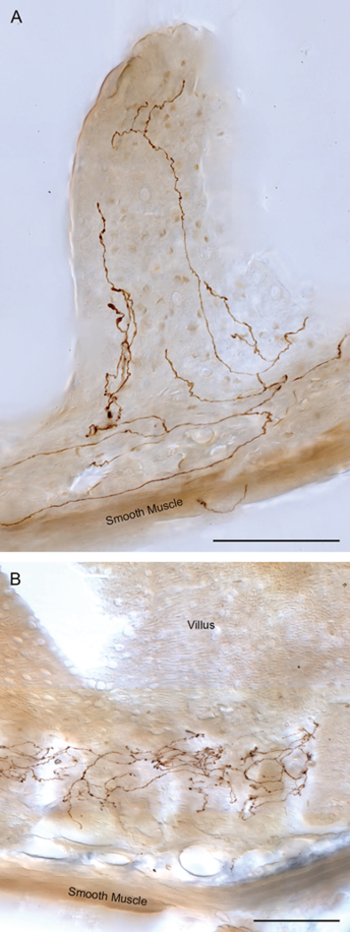
Brain-gut neural communications have long been considered limited because of conspicuous numerical mismatches. The vagus, the parasympathetic nerve connecting brain and gut, contains thousands of axons, whereas the gastrointestinal (GI) tract contains millions of intrinsic neurons in local plexuses. The numerical paradox was initially recognized in terms of efferent projections, but the number of afferents, which comprise the majority (≈ 80%) of neurites in the vagus, is also relatively small. The present survey of recent morphological observations suggests that vagal terminals, and more generally autonomic and visceral afferent arbors in the stomach as well as throughout the gut, elaborate arbors that are extensive, regionally specialized, polymorphic, polytopic, and polymodal, commonly with multiplicities of receptors and binding sites-smart terminals. The morphological specializations and dynamic tuning of one-to-many efferent projections and many-to-one convergences of contacts onto afferents create a complex architecture capable of extensive peripheral integration in the brain-gut connectome and offset many of the disparities between axon and target numbers. Appreciating this complex architecture can help in the design of therapies for GI disorders.

Keywords: afferent; autonomic; efferent; intestines; parasympathetic; postganglionic; preganglionic; stomach; vagus; viscera; visceral afferent.


Figures

Figure 1.
Figure 1.
Preganglionic projections to myenteric ganglia. (A) Simultaneous labeling of multiple motor projections (brown fibers: PHA-l labeling): large dorsal motor nucleus of vagus injections label efferent contacts on essentially all cells in the individual myenteric ganglia in the stomach (myenteric postganglionic neurons counterstained with Cuprolinic Blue). (B) Selective labeling of limited numbers of vagal preganglionic efferents with small injections (brown fibers: dextran-biotin labeling) reveal contacts around individual myenteric postganglionic neurons (individual postganglionic stained immunohistochemically for nNOS: steel gray secondary). (C) One of three nNOS-positive cells (steel gray secondary) selectively encircled by two preganglionic branches (brown fibers: dextran-biotin labeling). (D) Myenteric ganglia with immunohistochemistry for nNOS (steel gray positive postganglionic neurons) illustrate that some vagal preganglionic terminals selectively encircle the unstained (nNOS-negative) cells of the ganglion. Other preganglionic fibers (not shown; cf. panels B and C) preferentially contact nNOS-positive neurons within the myenteric plexus. Scale bars in plates = 30 μm (panel A), 16 μm (panel B), 25 μm (panel C), and 100 μm (panel D). Panel A reproduced by permission of John Wiley & Sons from Holst et al. Panel B reproduced by permission of Elsevier from Walter et al.
Figure 2.
Figure 2.
Digitization of individual vagal efferents establishes that single preganglionics link multiple neighboring myenteric ganglia and selectively innervate different phenotypes of myenteric postganglionic neurons. (A) Neurolucida® (MBF Bioscience, Williston, VT) reconstruction of whole mount and fiber location (upper left) and preganglionic arbor contacting multiple myenteric neurons in multiple ganglia (blue ganglia) within a larger field of ganglia un-innervated by the fiber (pale orange ganglia). Insets (B) and (C) illustrate the digitized vagal preganglionic fiber (dextran-biotin labeled) and its selectivity for certain myenteric neurons within the ganglia (counterstained with Cuprolinic blue). (D and E) In other specimens, immunohistochemistry used to selectively label nNOS neurons (steel gray chromogen) illustrates that some vagal preganglionic efferents (brown dextran-biotin labeled fibers) preferentially or selectively ring nNOS-negative or unstained (i.e., presumably cholinergic) profiles (e.g., panel D) whereas other efferents preferentially encircle nNOS-positive (presumably nitrergic) postganglionic neurons (e.g., panel E). Scale bars in plates = 1000 μm (panel A) and 25 μm (panels B−E).
Figure 3.
Figure 3.
Individual intraganglionic laminar endings (IGLEs) issued by a single afferent innervate multiple neighboring myenteric ganglia (red regions), apparently generating large receptive fields, but (unlike vagal efferents) IGLEs do not obviously discriminate or selectively innervate either nNOS-positive or -negative neurons within their receptive fields. (A) A Neurolucida reconstruction of a gastric whole mount and an IGLE afferent within it (right side of image) as well as an enlargement of the IGLE afferent (left part of figure). (B) A photomicrograph of a region of the IGLE afferent illustrated in (A), which reproduces several of the IGLE plates issued by the afferent as well as the myenteric ganglia (stained with a polyneuronal Cuprolinic blue protocol) that the afferent contacts. ( C, D, and E) Examples of IGLE plates from other specimens that illustrate the terminal patterns of IGLE afferents and display how they appear to contact local ensembles of adjacent neurons, whether they are nNOS-positive or -negative (panels C−E are counterstained by nNOS immunohistochemistry). Scale bars in plates = 500 μm (panel A), 250 μm (panel B), and 25 μm (panels C−E).
Figure 4.
Figure 4.
The conventional IGLE phenotype exhibits regional specializations. IGLE branches, particularly in the distal stomach, often have more varicose, less lamelliform specializations of puncta. (A’) Inset illustrates common IGLE puncta morphology of flattened lamelliform puncta, frequently displaying spinous extensions, routinely observed in the proximal stomach. (A, B, and C) In the distal stomach, IGLE afferents often issue some—or even a majority of—branches that terminate in rounded or beaded varicosities, rather than flattened and lamelliform puncta. These IGLE specializations can consist of branches with a mixture of varicosities and smaller lamelliform puncta (e.g., panel A), branches issuing almost exclusively simpler varicosities (e.g., panel B), or branches terminating in dense aggregations of simple and varicose puncta (e.g., panel C). Scale bars = 10 μm (panel A’), 25 μm (panels A and B), and 50 μm (panel C).
Figure 5.
Figure 5.
Intramuscular arrays (IMAs), in contrast to vagal preganglionic efferents and IGLE afferents, directly innervate the muscle layers and form arrays of branches that typically run with the ICC network found in the smooth muscle layers. Panels A, B, and C are photomicrographs of branches of the IMA digitized with Neurolucida in panel E. Panel D illustrates how IMA branches (dextran-biotin labeled brown fiber) course with ICCs (elongated purple cell—Vector® VIP labeled for cKit antibody) in the muscle layers. Panel F illustrates the specialized “web ending” variant of IMAs seen near the distal antral/pyloric insertion of sling muscle fibers. Panel G is a higher power image of the IMA apparatus and its contacts or varicosities illustrated in panel F. Scale bars = 20 μm (panels A−C), 10 μm (panel D), 250 μm (panels E and F), and 25 μm (panel G). Panels A–E reproduced by permission of Elsevier from Powley and Phillips.
Figure 6.
Figure 6.
Vagal mucosal arbors are afferents that arborize deep in the mucosal layer and send a subset of their branches paralleling the gastric glands and reaching the basal side of the epithelial wall in direct contact with the contents of the stomach. Panel A illustrates a dextranbiotin filled mucosal arbor (brown branches) in the mucosal layer. Panel B illustrates how the mucosal fibers branch and ramify in the deeper mucosal layers in the zone heavily populated with EEC (in the specimen in panel B, the brown dextran-biotin labeled afferent articulates with EEC immunohistochemically stained for gastrin (steel gray secondary)). Scale bars = 100 μm.
Figure 7.
Figure 7.
Vagal afferents innervate villi and crypts (or intestinal glands) throughout the small intestines. Panel A illustrates a villus ending (brown neurites labeled with dextran-biotin) in the distal jejunum. Vagal villus afferents throughout the small intestine issue multiple branches that course along the basal side of the epithelial wall and run apically to the villus tips. Several adjacent villi can be innervated by one afferent arbor. Along the small intestine, the vagal villus afferents exhibit local specializations with, generally, more numerous branches in individual villi in the proximal intestines and less numerous branches in the distal intestines. Panel B illustrates the arbor of a villus crypt—or gland—afferent (brown neurites labeled with dextran-biotin) encircling multiple neighboring glands immediately below intestinal villi. This afferent, located in the distal duodenum, characteristically links several neighboring glands into a presumptive receptive field. Scale bars = 100 μm.
Figure 8.
Figure 8.
Sympathetic efferent fibers innervating the gut exhibit complex terminal arbors. The labeled fiber in this photomicrograph (brown neurite labeled with dextran biotin) courses through and contacts myenteric ganglion cells (entering at lower right) and then turns and continues to extensively arborize in the smooth muscle wall, where it produces branches coursing both longitudinally (top to bottom of figure) and circularly (left to right of figure). Scale bar = 31 μm. Reproduced by permission of John Wiley & Sons from Walter et al.

References

    1. Langley JN 1922. Connexions of the enteric nerve cells. J. Physiol. (Lond.). 56:xxxix
    1. Wood JD 1987. Physiology of the Enteric Nervous System In Physiology of the Gastrointestinal Tract. Johnson LR, Ed.: 2nd ed. New York, Raven Press.
    1. Hill CJ 1927. A contribution to our knowledge of the enteric plexuses. Philos. Trans. R. Soc. Lond. B Biol. Sci. 215:355–387.
    1. Berthoud H-R, Jedrzejewska A, & Powley TL. 1990. Simultaneous labeling of vagal innervation of the gut and afferent projections from the visceral forebrain with Dil injected into the dorsal vagal complex in the rat. J. Comp. Neurol. 301(1):65–79. - PubMed
    1. Holst M-C, Kelly JB & Powley TL. 1997. Vagal preganglionic projections to the enteric nervous system characterized with PHA-L. J. Comp. Neurol. 381:81–100. - PubMed

Publication types