Review
. 2019 Jul 1;38(13):e100278.
doi: 10.15252/embj.2018100278. Epub 2019 May 14.

Uncovering the assembly pathway of human ribosomes and its emerging links to disease

Affiliations
Review

Uncovering the assembly pathway of human ribosomes and its emerging links to disease

Katherine E Bohnsack et al. EMBO J. .

Abstract

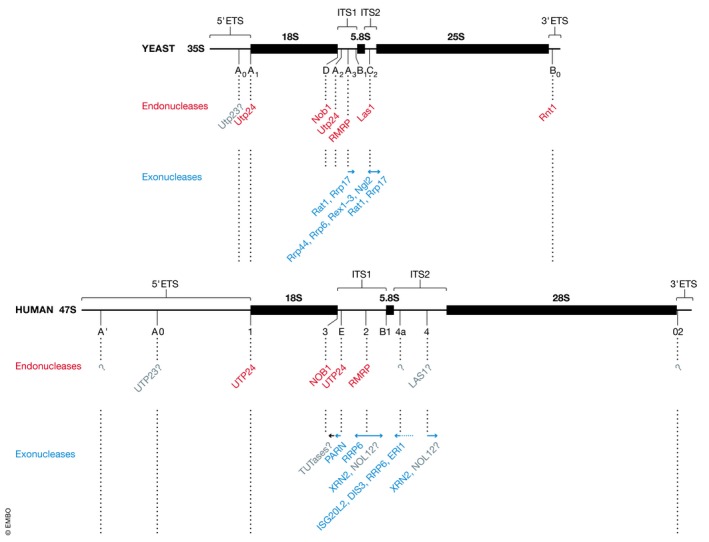
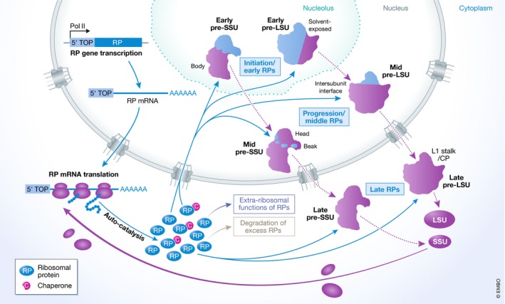
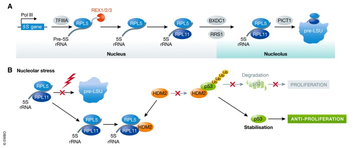
The essential cellular process of ribosome biogenesis is at the nexus of various signalling pathways that coordinate protein synthesis with cellular growth and proliferation. The fact that numerous diseases are caused by defects in ribosome assembly underscores the importance of obtaining a detailed understanding of this pathway. Studies in yeast have provided a wealth of information about the fundamental principles of ribosome assembly, and although many features are conserved throughout eukaryotes, the larger size of human (pre-)ribosomes, as well as the evolution of additional regulatory networks that can modulate ribosome assembly and function, have resulted in a more complex assembly pathway in humans. Notably, many ribosome biogenesis factors conserved from yeast appear to have subtly different or additional functions in humans. In addition, recent genome-wide, RNAi-based screens have identified a plethora of novel factors required for human ribosome biogenesis. In this review, we discuss key aspects of human ribosome production, highlighting differences to yeast, links to disease, as well as emerging concepts such as extra-ribosomal functions of ribosomal proteins and ribosome heterogeneity.

Keywords: RNA modification; RNA processing; ribosomal protein; ribosome; ribosomopathy.


Conflict of interest statement

The authors declare that they have no conflict of interest.

Figures

Figure 1
Figure 1. Pre‐rRNA processing in yeast and human cells
Schematic views of the primary pre‐rRNA transcripts from yeast (35S, upper panel) and human cells (47S, lower panel) are shown to scale. Mature 18S, 5.8S and 25S/28S rRNA sequences are indicated by black rectangles, external transcribed spacers (5′‐ETS and 3′‐ETS) and internal transcribed spacers (ITS1 and ITS2) are represented by black lines. Relative cleavage site positions are indicated, and sites are specified below the pre‐rRNA transcripts. Enzymes responsible for individual processing steps are indicated (Endonucleases—red; exonucleases—blue; putative enzymes—grey; unknown enzymes—?).
Figure 2
Figure 2. Distribution of modifications in human rRNAs
Tertiary structures of the human small ribosomal subunit (SSU) and large ribosomal subunit (LSU; PBD 4V6X) are shown, with rRNA sequences depicted as ribbons and ribosomal proteins in surface view. The type and nucleotide position of base modifications, as well as the positions of key functional regions of the ribosome, are indicated.
Figure 3
Figure 3. Production, recruitment and functions of human ribosomal proteins
Schematic model showing the expression and assembly of ribosomal proteins (RP). Red arrows indicated sequential steps in the process, and dashed black lines indicate maturation of pre‐ribosomal particles. Pink circles containing a white “C” depict dedicated ribosomal protein chaperones. CP, central protuberance.
Figure 4
Figure 4. Assembly of the 5S RNP and its role in regulation of the tumour suppressor p53
(A) Model of synthesis and assembly of the 5S RNP from 5S rRNA and ribosomal proteins RPL5 (uL18) and RPL11 (uL5), and 5S RNP integration into the pre‐LSU. (B) Upon nucleolar stress (e.g. in ribosomopathies), 5S RNP integration into the pre‐LSU is decreased and 5S RNP accumulates in the nucleoplasm, where it binds the E3 ubiquitin ligase HDM2. This impedes association of HDM2 with p53, promoting p53 stabilisation and inhibiting cell proliferation.

References

    1. Ameismeier M, Cheng J, Berninghausen O, Beckmann R (2018) Visualizing late states of human 40S ribosomal subunit maturation. Nature 558: 249–253 - PubMed
    1. Andersen JS, Lyon CE, Fox AH, Leung AKL, Lam YW, Steen H, Mann M, Lamond AI (2002) Directed proteomic analysis of the human nucleolus. Curr Biol 12: 1–11 - PubMed
    1. Anger AM, Armache J‐P, Berninghausen O, Habeck M, Subklewe M, Wilson DN, Beckmann R (2013) Structures of the human and Drosophila 80S ribosome. Nature 497: 80–85 - PubMed
    1. Ansel KM, Pastor WA, Rath N, Lapan AD, Glasmacher E, Wolf C, Smith LC, Papadopoulou N, Lamperti ED, Tahiliani M et al (2008) Mouse Eri1 interacts with the ribosome and catalyzes 5.8S rRNA processing. Nat Struct Mol Biol 15: 523–530 - PMC - PubMed
    1. Armistead J, Khatkar S, Meyer B, Mark BL, Patel N, Coghlan G, Lamont RE, Liu S, Wiechert J, Cattini PA et al (2009) Mutation of a gene essential for ribosome biogenesis, EMG1, causes bowen‐conradi syndrome. Am J Hum Genet 84: 728–739 - PMC - PubMed

Publication types

Substances

LinkOut - more resources