

Periodontal bacterial supernatants modify differentiation, migration and inflammatory cytokine expression in human periodontal ligament stem cells
- PMID: 31269072
- PMCID: PMC6609032
- DOI: 10.1371/journal.pone.0219181
Periodontal bacterial supernatants modify differentiation, migration and inflammatory cytokine expression in human periodontal ligament stem cells
Abstract
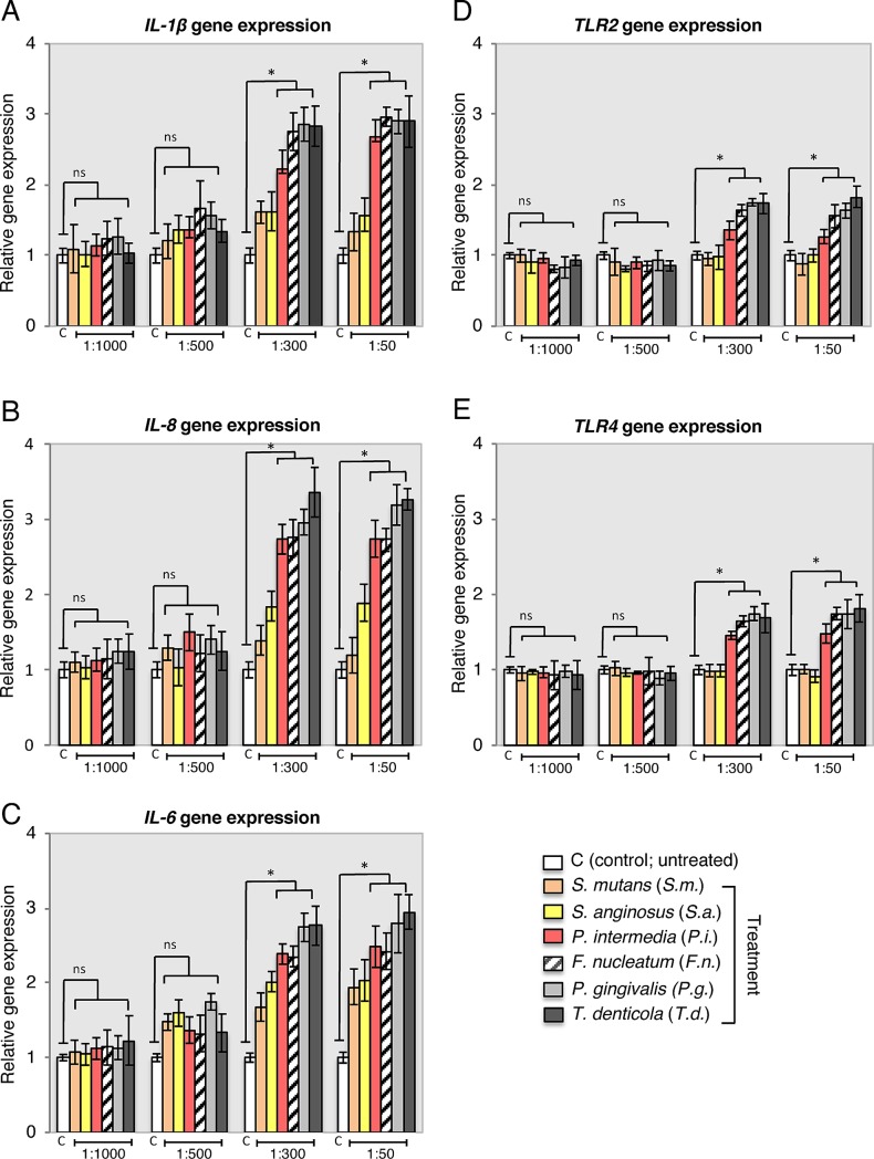
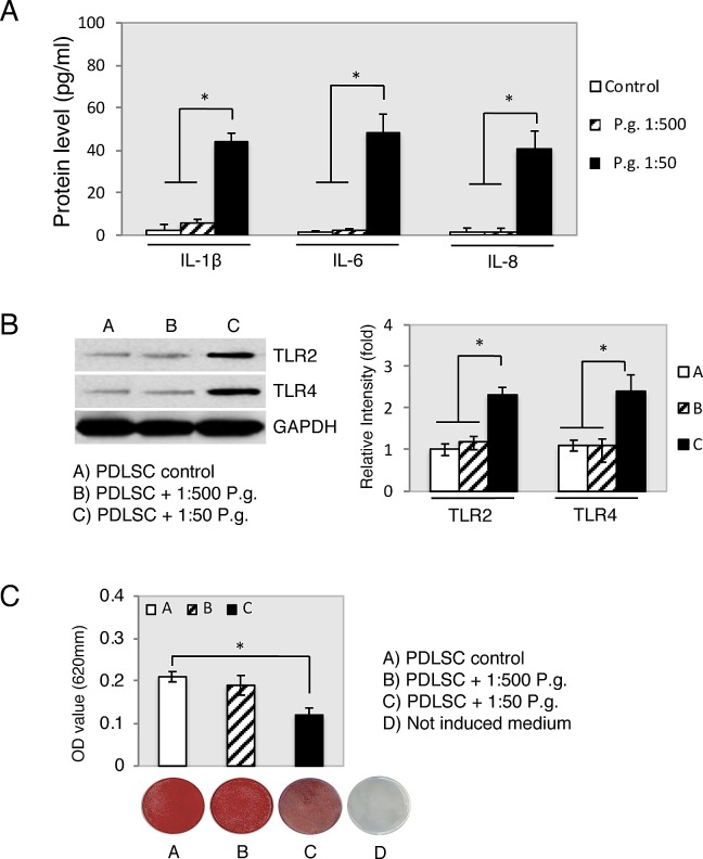
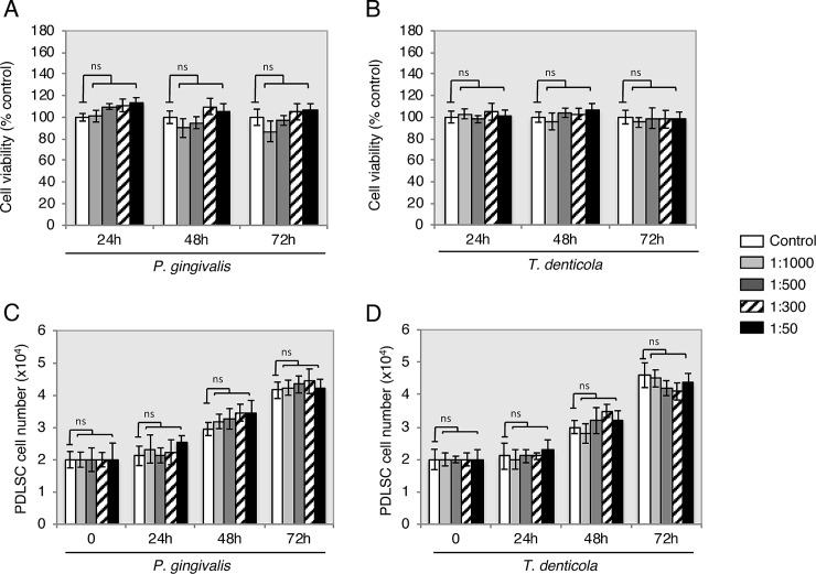
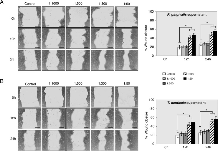
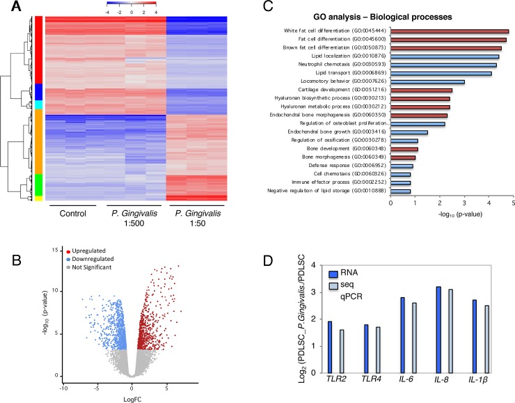
Periodontal ligament stem cells (PDLSC) play an important role in periodontal tissue homeostasis/turnover and could be applied in cell-based periodontal regenerative therapy. Bacterial supernatants secreted from diverse periodontal bacteria induce the production of cytokines that contribute to local periodontal tissue destruction. However, little is known about the impact of whole bacterial toxins on the biological behavior of PDLSC. Therefore this study investigated whether proliferation, migration, inflammatory cytokines expression and transcriptional profile would be affected by exposure to endotoxins from bacterial species found in the subgingival plaque. PDLSC were cultured with the following bacterial supernatants: S. mutans, S. anginosus, P. intermedia, F. nucleatum, P. gingivalis and T. denticola. These supernatants were prepared in dilutions of 1:1000, 1:500, 1:300 and 1:50. Using quantitative RT-PCR, gene expression of selected inflammatory cytokines (IL-6, IL-8 and IL-1β) and cell-surface receptors (TLR2, TLR4) showed upregulation of ≈2.0- to 3.0-fold, when exposed to P. intermedia, F. nucleatum, P. gingivalis and T. denticola. However, supernatants did not affect proliferation (MTT) and migration (wound scratch assays) of PDLSC. Next generation RNA sequencing confirmed modified lineage commitment of PDLSC by stimulating chondrogenesis, adipogenesis and inhibition of osteogenesis under P. gingivalis supernatant treatment compared to control. Taken together, this study shows stem cell immunomodulatory response to different periodontal bacteria supernatant and suggests that stem cell transcriptional capacity, migration/proliferation and osteogenesis may differ in the presence of those pathogens. These results bring into question stem cell contribution to periodontal tissue regeneration and onset of inflammation.
Conflict of interest statement
The authors have declared that no competing interests exist.
Figures
Similar articles
-
[Effect of trichostatin A on the osteogenic differentiation potential of periodontal ligament stem cells in inflammatory microenvironment induced by tumor necrosis factor-α stimulation].Zhonghua Kou Qiang Yi Xue Za Zhi. 2016 Apr 9;51(4):235-41. doi: 10.3760/cma.j.issn.1002-0098.2016.04.010. Zhonghua Kou Qiang Yi Xue Za Zhi. 2016. PMID: 27117217 Chinese.
-
Lipopolysaccharide can modify differentiation and immunomodulatory potential of periodontal ligament stem cells via ERK1,2 signaling.J Cell Physiol. 2018 Jan;233(1):447-462. doi: 10.1002/jcp.25904. Epub 2017 May 19. J Cell Physiol. 2018. PMID: 28295277
-
Osmunda japonica Extract Suppresses Pro-Inflammatory Cytokines by Downregulating NF-κB Activation in Periodontal Ligament Fibroblasts Infected with Oral Pathogenic Bacteria.Int J Mol Sci. 2020 Apr 1;21(7):2453. doi: 10.3390/ijms21072453. Int J Mol Sci. 2020. PMID: 32244806 Free PMC article.
-
Periodontal Ligament Stem Cells: Current Knowledge and Future Perspectives.Stem Cells Dev. 2019 Aug 1;28(15):995-1003. doi: 10.1089/scd.2019.0025. Epub 2019 May 20. Stem Cells Dev. 2019. PMID: 31017047 Review.
-
Non-coding RNAs Function in Periodontal Ligament Stem Cells.Stem Cell Rev Rep. 2024 Aug;20(6):1521-1531. doi: 10.1007/s12015-024-10731-5. Epub 2024 Jun 7. Stem Cell Rev Rep. 2024. PMID: 38848014 Review.
Cited by
-
Baricitinib alleviates lipopolysaccharide‑induced human periodontal ligament stem cell injury and promotes osteogenic differentiation by inhibiting JAK/STAT signaling.Exp Ther Med. 2022 Dec 22;25(2):74. doi: 10.3892/etm.2022.11773. eCollection 2023 Feb. Exp Ther Med. 2022. PMID: 36684656 Free PMC article.
-
Unveiling the Differences in Biological Properties of Dental Pulp Stem Cells from Normal and Inflamed Pulp: A Comprehensive Comparative Study.Med Sci Monit. 2022 Mar 18;28:e934511. doi: 10.12659/MSM.934511. Med Sci Monit. 2022. PMID: 35301274 Free PMC article.
-
Bacterial growth stage determines the yields, protein composition, and periodontal pathogenicity of Porphyromonas gingivalis outer membrane vesicles.Front Cell Infect Microbiol. 2023 Oct 11;13:1193198. doi: 10.3389/fcimb.2023.1193198. eCollection 2023. Front Cell Infect Microbiol. 2023. PMID: 37900318 Free PMC article.
-
Levels of Gene Expression of Immunological Biomarkers in Peri-Implant and Periodontal Tissues.Int J Environ Res Public Health. 2020 Dec 6;17(23):9100. doi: 10.3390/ijerph17239100. Int J Environ Res Public Health. 2020. PMID: 33291232 Free PMC article.
-
Propolis, Aloe Vera, Green Tea, Cranberry, Calendula, Myrrha and Salvia Properties against Periodontal Microorganisms.Microorganisms. 2022 Oct 31;10(11):2172. doi: 10.3390/microorganisms10112172. Microorganisms. 2022. PMID: 36363764 Free PMC article. Review.
References
-
- Page RC & Kornman KS. The pathogenesis of human periodontitis: an introduction. Periodontology 2000. 1997; 14: 9–11. - PubMed
MeSH terms
Substances
LinkOut - more resources
Full Text Sources
Medical
