. 2019 Nov;34(11):2117-2132.
doi: 10.1002/jbmr.3825. Epub 2019 Oct 9.

Regulation of the Bone Vascular Network is Sexually Dimorphic

Affiliations

Regulation of the Bone Vascular Network is Sexually Dimorphic

Alice Goring et al. J Bone Miner Res. 2019 Nov.

Abstract

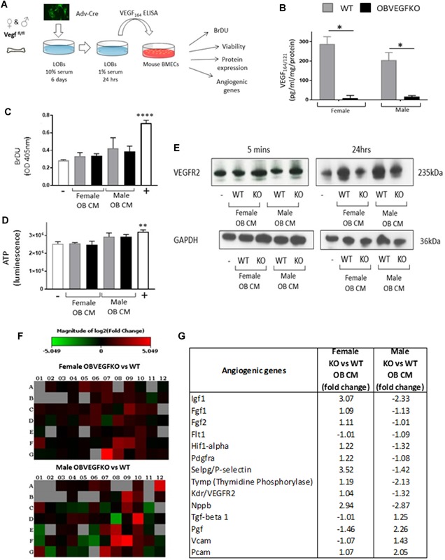
Osteoblast (OB) lineage cells are an important source of vascular endothelial growth factor (VEGF), which is critical for bone growth and repair. During bone development, pubertal differences in males and females exist, but little is known about whether VEGF signaling contributes to skeletal sexual dimorphism. We have found that in mice, conditional disruption of VEGF in osteocalcin-expressing cells (OcnVEGFKO) exerts a divergent influence on morphological, cellular, and whole bone properties between sexes. Furthermore, we describe an underlying sexual divergence in VEGF signaling in OB cultures in vitro independent of circulating sex hormones. High-resolution synchrotron computed tomography and backscattered scanning electron microscopy revealed, in males, extensive unmineralized osteoid encasing enlarged blood vessel canals and osteocyte lacunae in cortical bone after VEGF deletion, which contributed to increased porosity. VEGF was deleted in male and female long bone-derived OBs (OBVEGKO) in vitro and Raman spectroscopic analyses of mineral and matrix repertoires highlighted differences between male and female OBVEGFKO cells, with increased immature phosphate species prevalent in male OBVEGFKO cultures versus wild type (WT). Further sexual dimorphism was observed in bone marrow endothelial cell gene expression in vitro after VEGF deletion and in sclerostin protein expression, which was increased in male OcnVEGFKO bones versus WT. The impact of altered OB matrix composition after VEGF deletion on whole bone geometry was assessed between sexes, although significant differences between OcnVEGFKO and WT were identified only in females. Our results suggest that bone-derived VEGF regulates matrix mineralization and vascularization distinctly in males and females, which results in divergent physical bone traits.

Keywords: BONE QCT/MICROCT; GENETIC ANIMAL MODELS; MATRIX MINERALIZATION; OSTEOBLASTS; PRECLINICAL STUDIES.


Figures

Figure 1
Figure 1
Conditional deletion of VEGF alters bone formation in adult tibiae. No gross anatomical differences were evident between OcnVEGFKO versus WT animals at 16 weeks of age (A; scale bar = 1 cm). Similarly, tail length (cm) was not significantly different between OcnVEGFKO and WT groups (B). No significant differences in the weight (g) of male OcnVEGFKO versus WT were observed (C) (B, C, n = 6–8 animals from 5 individual litters, presented as mean measurements ±SEM *p < 0.05, ****p < 0.0001 using two‐way ANOVA). Whole bone scans (D; 18 μm resolution) revealed poorly mineralized regions of the OcnVEGFKO tibia localized to the epiphysis and the tibiofibular junction (arrows) specifically in male animals. Tibial length (mm) was significantly greater in male OcnVEGFKO than WT (E; n = 4 males and 4 females from individual litters presented as mean ±SEM *p < 0.05 using two‐way ANOVA). Total volume porosity (F) was calculated after reconstruction of 300 SRCT slices lacunae (error bars indicate mean value ±SEM, n = 3 females and 3 males from individual litters ****p < 0.0001 using two‐way ANOVA). High‐resolution, synchrotron X‐ray computed tomography (SRCT) (G; 0.65 μm) slices from female and male WT and OcnVEGFKO mice revealed poorly mineralized areas of cortical bone at posterior region of tibiofibular junction (G; white arrows) and differences in cortical porosity between male and female OcnVEGFKO. Scale bar = 200 µm.
Figure 2
Figure 2
Distinction of bone porosity components is compromised after VEGF deletion in males. 3D renderings of osteocyte lacunae (yellow) and intracortical canals (red) from female (A) and male (B) WT and OcnVEGFKO mice (16 weeks old) after SRCT scans (0.65 μm voxel size). Scale bar = 50 µm. In WT male and female animals, osteocyte lacunae made up the highest fraction of the intracortical porosity (C). In male OcnVEGFKO, % pores constituting the intracortical canal fraction was increased versus WT (D). Measurements taken of regularly sized osteocyte lacunae show an increase in lacuna number density (Lc.Dn; E), mean lacunar volume (Lc.V; F), and mean lacunar diameter (Lc.Dm; G) in female OBVEGFKO versus WT. In males, there was a decrease in mean lacunar diameter (Lc.Dm; G) after knockout of OBVEGF. The probability density of osteocytes of different volumes for females (H) and males (I) are shown by graphs on a logarithmic scale, which were created using the free language and environment for statistical computing and graphics R (https://cran.r-project.org/). Each colored line represents one animal. Number of intracortical canals greater than the lacunar threshold (see Materials and Methods) is significantly decreased in male OcnVEGFKO (Ca.Dn; J). An increase in mean canal volume was observed after OcnVEGFKO (Ca.V; K); however, no change in mean canal diameter was observed in male OcnVEGFKO (Ca.Dm; L). Error bars indicate mean value ±SEM, n = 3 females and 3 males from individual litters *p < 0.05, **p < 0.01, ***p < 0.001, ****p < 0.0001 using two‐way ANOVA.
Figure 3
Figure 3
Conditional VEGF deletion increases blood vessel area and unmineralized osteoid in males. No obvious differences in bone architecture are found between female WT (A) and OcnVEGFKO bones (B) in either the SEM images (left; scale bar = 100 µm) or the pentachrome‐stained bone sections (right; scale bar = 20 µm). SEM shows very few blood vessels remaining in both the WT and OcnVEGFKO after treatment with HCl and NaOCL. There are minimal areas of osteoid surrounding the BV canals and osteocyte lacunae. In comparison to male WT (C), in OcnVEGFKO bones (D), increased vascularization (red arrows) was present and visible in SEM images after treatment with HCl and NaOCL (left) and in pentachrome‐stained images (right). The close proximity and overlapping of the osteocytes surrounding the bone marrow (BM) and the poor mineralization result in large amounts of PMMA remaining. In histologically stained male OcnVEGFKO sections, there were large amounts of unmineralized osteoid (*) surrounding BV canals (hatched lines). Analysis of stained sections showed that OcnVEGFKO versus WT % area BV canals (E) was increased in males. There was a high level of vascular canal occupancy (F), which remained unchanged in OcnVEGFKO versus WT animals (error bars indicate mean value ±SEM, n = 4 female and n = 3 male mice from individual litters, **p < 0.01 using two‐way ANOVA). Raman spectroscopy revealed a lack of hydroxyapatite (HA) in perivascular regions versus cortical bone (G).
Figure 4
Figure 4
Sexually dimorphic effects of VEGF deletion on endothelial cell function. Mouse bone marrow endothelial cells (MBMECs) were treated with LOB conditioned media (CM) from male and female WT and OBVEGFKO cells for 24 hours (A). VEGF ELISA confirms VEGF deletion (B; three separate experiments ±SEM, *p < 0.05 using t test). For proliferation (BrDU; C) and viability (ATP; D) assays, MBMECs were treated for 24 hours with WT or OBVEGFKO CM with low‐serum (1%) media (–) and high serum (10%; +) used as controls. Data represent n = 6 replicates ±SEM, **p < 0.01, ****p < 0.0001 using t test. For VEGFR2 expression, MBMECs were treated for either 5 minutes or 24 hours and total VEGFR2 protein assessed by Western blotting (E). Gene expression changes were assessed by endothelial qPCR array with heat maps representing expression changes (fold change) of 84 different genes after the treatment of MBMEC with male and female OBVEGFKO and WT conditioned media (F). Table summarizes changes in gene expression in both sexes (G).
Figure 5
Figure 5
Direct effects of VEGF deletion on male and female osteoblast function. VEGF was deleted in vitro in male and female LOBs (P4). No differences in viability (ATP; A) versus WT were evident from males or females. Control cells received low‐serum media (–) Wnt3a (75 ng/mL +). Data represent mean value from six individual infections ±SEM, ***p < 0.001, ****p < 0.0001 using t test. Significant increases were found in alkaline phosphatase (B) after OBVEGFKO (C; scale bar = 100 µm). Control cells received 10% FBS media (–) and 10% FBS containing β‐glycerol‐phosphate (+). Data represent mean value from three individual infections ±SEM, **p < 0.01, ***p < 0.001 using t test. Knock‐down efficiency was further confirmed using qPCR (D) for VEGF mRNA in male and female OBVEGFKOs. The involvement of osteoclasts in the phenotype was investigated looking at relative expression of OPG (E) and RANKL (F), where no significant changes were identified after OBVEGFKO. Raman spectroscopy was able to detect clear and significant sex differences (between immature (ACP; G), intermediate (OCP; H), and mature (CAP; I) calcium species in WT and OBVEGFKO cells. Mineral/matrix ratio also shows a significant increase in matrix maturity after OBVEGFKO in females, whereas the converse is exhibited in males (J). Schematic representation of the changes in calcium phosphate species in WT and OBVEGFKO cells are shown (K). Error bars indicate mean value ±SEM, **p < 0.01, ****p < 0.0001 using t test, n = 50 spectra from each treatment group. Cell number increased over time in culture (****p < 0.0001) in both sexes but was not affected by OBVEGFKO (L). Data represent mean number of nuclei per µm2 ±SEM, ****p < 0.0001 using two‐way ANOVA, n = 20 fields of view per group. No notable changes in gene expression of androgen receptor (Ar; M), estrogen receptor 1 (Esr1; N), or estrogen receptor 2 (Esr2; O) were evident in males and females after OBVEGFKO.
Figure 6
Figure 6
Sexual dimorphic alterations in protein expression of SOST after VEGF deletion are evident in whole bone sections. Cryosections from female (A) and male (B) WT and OcnVEGFKO tibiofibular junction were stained with sclerostin primary antibody and Hoechst to stain the nuclei. Increases in sclerostin levels were visible after OcnVEGFKO in males (Alexa Fluor 555), specifically in the posterior region (arrows) below the bone marrow (BM). White box represents the region of cortical bone magnified below. Scale bar = 100 µm.
Figure 7
Figure 7
Significant alterations in tibial geometry after bone‐derived VEGF deletion is evident only in females. Minimum and maximum second moments of inertia (Imin and Imax, respectively), cross‐sectional area, resistance to torsion (J), ellipticity, cortical thickness, and bone mineral density (BMD) of male and female OcnVEGFKO tibiae versus WT at 16 weeks of age (A). Graphical heat map summarizes statistical differences (using ANOVA) at specific matched locations along the tibial length (10% to 90%), representative of overall effect of genotype. Red p ≤ 0.0001, yellow p ≤ 0.001–0.01, green p ≤ 0.01–0.05, and blue p ≥ 0.05. Line graphs represent means for female (B) and male (C) WT versus OcnVEGFKO ±SEM (n = 4 female and 4 male mice from individual litters). Longitudinal tibial cross sections created using CTvox show a decrease in bone density in OcnVEGFKO females (D) and an increase in bone density in OcnVEGFKO males (E). Heat map scale plotted from 0 (brown; low‐threshold density) to 255 (blue; high‐threshold density).
Figure 8
Figure 8
Sexually dimorphic effects of VEGF deletion on matrix mineralization and porosity link to whole bone phenotypes. Female OcnVEGFKO mice are able to maintain porosity and vasculature but compromise tibial geometry after deletion of VEGF in osteoblast cells. In contrast, male OcnVEGFKOs are able to maintain tibial geometry, despite increased cortical porosity associated with altered blood vessel configuration. Raman spectroscopy has shown that this skeletal sexual dimorphism could be linked to direct effects of VEGF on OB matrix production, with increased immature phosphate species (ACP) predominant in male OBVEGFKO cells.

Similar articles

Cited by

References

    1. Liu Y, Olsen BR. Distinct VEGF functions during bone development and homeostasis. Arch Immunol Ther Exp (Warsz). 2014;62:363–8. - PubMed
    1. Kanczler JM, Oreffo RO. Osteogenesis and angiogenesis: the potential for engineering bone. Eur Cell Mater. 2008;15:100–14. - PubMed
    1. Clarkin CE, Gerstenfeld LC. VEGF and bone cell signaling: an essential vessel for communication? Cell Biochem Funct. 2013;31:1–11. - PubMed
    1. Hu K, Olsen BR. The roles of vascular endothelial growth factor in bone repair and regeneration. Bone. 2016;91:30–8. - PMC - PubMed
    1. Liu Y, Berendsen AD, Jia S, et al. Intracellular VEGF regulates the balance between osteoblast and adipocyte differentiation. J Clin Invest. 2012;122:3101–13. - PMC - PubMed

Publication types

Substances