

Neuroanatomy Learning: Augmented Reality vs. Cross-Sections
- PMID: 31269322
- PMCID: PMC7317366
- DOI: 10.1002/ase.1912
Neuroanatomy Learning: Augmented Reality vs. Cross-Sections
Abstract
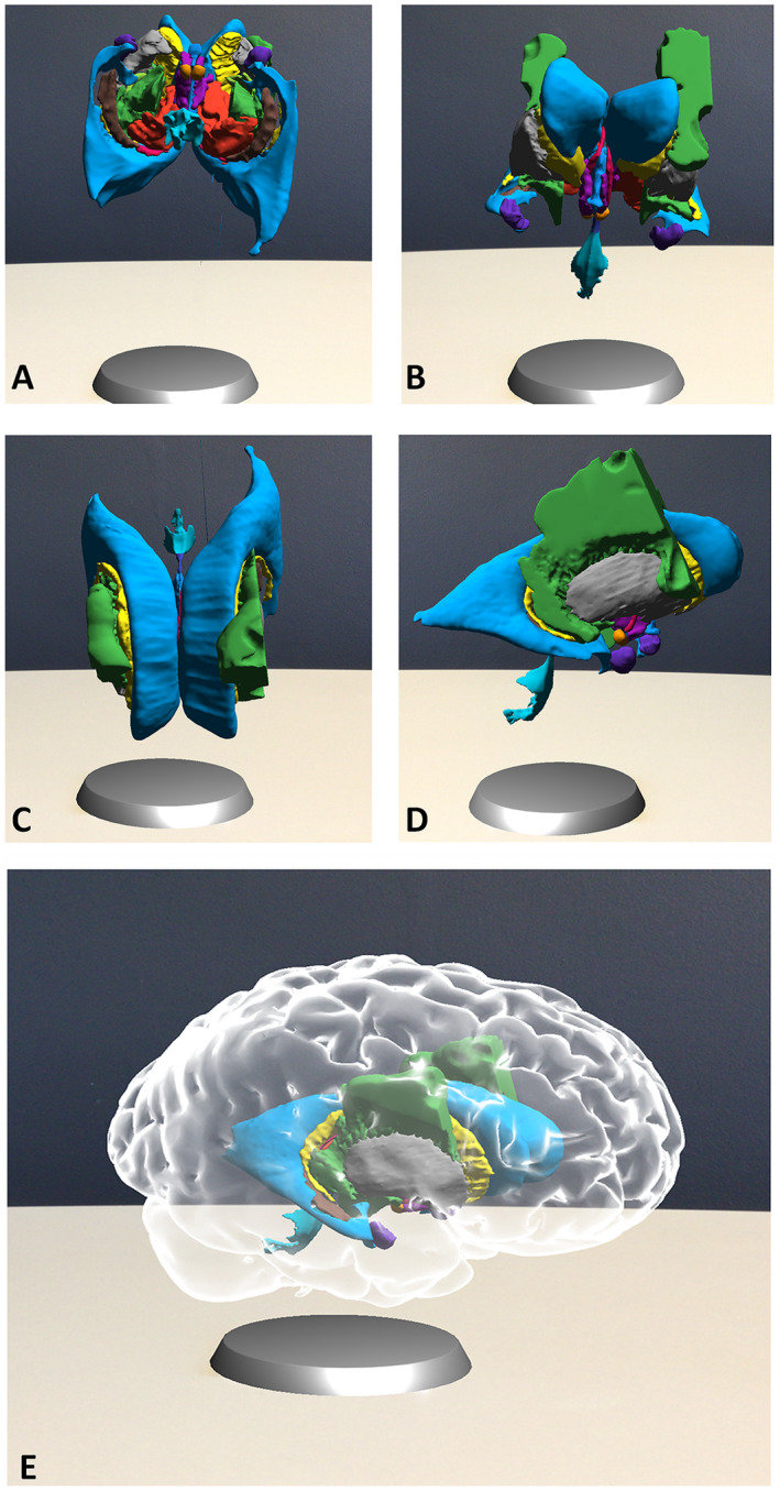
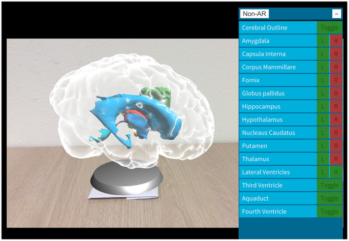
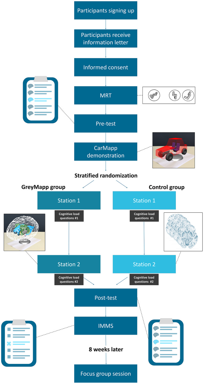
Neuroanatomy education is a challenging field which could benefit from modern innovations, such as augmented reality (AR) applications. This study investigates the differences on test scores, cognitive load, and motivation after neuroanatomy learning using AR applications or using cross-sections of the brain. Prior to two practical assignments, a pretest (extended matching questions, double-choice questions and a test on cross-sectional anatomy) and a mental rotation test (MRT) were completed. Sex and MRT scores were used to stratify students over the two groups. The two practical assignments were designed to study (1) general brain anatomy and (2) subcortical structures. Subsequently, participants completed a posttest similar to the pretest and a motivational questionnaire. Finally, a focus group interview was conducted to appraise participants' perceptions. Medical and biomedical students (n = 31); 19 males (61.3%) and 12 females (38.7%), mean age 19.2 ± 1.7 years participated in this experiment. Students who worked with cross-sections (n = 16) showed significantly more improvement on test scores than students who worked with GreyMapp-AR (P = 0.035) (n = 15). Further analysis showed that this difference was primarily caused by significant improvement on the cross-sectional questions. Students in the cross-section group, moreover, experienced a significantly higher germane (P = 0.009) and extraneous cognitive load (P = 0.016) than students in the GreyMapp-AR group. No significant differences were found in motivational scores. To conclude, this study suggests that AR applications can play a role in future anatomy education as an add-on educational tool, especially in learning three-dimensional relations of anatomical structures.
Keywords: augmented reality; cross-sectional anatomy; medical education; neuroanatomy; neuroanatomy education; neuroscience; undergraduate education.
© 2019 The Authors. Anatomical Sciences Education published by Wiley Periodicals, Inc. on behalf of American Association of Anatomists.
Figures

Comment in
-
Letter to the Editor. Considerations for the value of extended reality versus ex cathedra format for neuroanatomy education.Neurosurg Focus. 2024 Jun;56(6):E19. doi: 10.3171/2024.2.FOCUS2485. Neurosurg Focus. 2024. PMID: 38823048 No abstract available.
References
-
- Albanese M. 2010. The gross anatomy laboratory: A prototype for simulation‐based medical education. Med Educ 44:7–9. - PubMed
-
- Allen LK, Eagleson R, de Ribaupierre S. 2016. Evaluation of an online three‐dimensional interactive resource for undergraduate neuroanatomy education. Anat Sci Educ 9:431–439. - PubMed
-
- Anglin GJ, Vaez H, Cunningham KL. 2004. Visual representations and learning: The role of static and animated graphics In: Jonassen DH. (Editor). Handbook of Research on Educational Communications and Technology. 2nd Ed. Mahwah, NJ: Lawrence Erlbaum Associates Inc., Publishers; p 865–916.
-
- Arnts H, Kleinnijenhuis M, Kooloos JG, Schepens‐Franke AN, van Cappellen van Walsum AM. 2014. Combining fiber dissection, plastination, and tractography for neuroanatomical education: Revealing the cerebellar nuclei and their white matter connections. Anat Sci Educ 7:47–55. - PubMed
Publication types
MeSH terms
Grants and funding
LinkOut - more resources
Full Text Sources
Research Materials
