Review
. 2019 Jul 2;30(1):51-65.
doi: 10.1016/j.cmet.2019.05.023.

GPIHBP1 and Lipoprotein Lipase, Partners in Plasma Triglyceride Metabolism

Affiliations
Review

GPIHBP1 and Lipoprotein Lipase, Partners in Plasma Triglyceride Metabolism

Stephen G Young et al. Cell Metab. .

Abstract

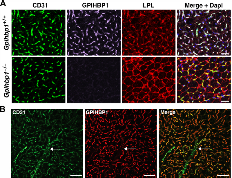
Lipoprotein lipase (LPL), identified in the 1950s, has been studied intensively by biochemists, physiologists, and clinical investigators. These efforts uncovered a central role for LPL in plasma triglyceride metabolism and identified LPL mutations as a cause of hypertriglyceridemia. By the 1990s, with an outline for plasma triglyceride metabolism established, interest in triglyceride metabolism waned. In recent years, however, interest in plasma triglyceride metabolism has awakened, in part because of the discovery of new molecules governing triglyceride metabolism. One such protein-and the focus of this review-is GPIHBP1, a protein of capillary endothelial cells. GPIHBP1 is LPL's essential partner: it binds LPL and transports it to the capillary lumen; it is essential for lipoprotein margination along capillaries, allowing lipolysis to proceed; and it preserves LPL's structure and activity. Recently, GPIHBP1 was the key to solving the structure of LPL. These developments have transformed the models for intravascular triglyceride metabolism.

Keywords: chylomicronemia; endothelial cells; hypertriglyceridemia; lipid transport; lipoprotein lipase.


Figures

Figure 1
Figure 1. A cartoon representation of GPIHBP1’s functions in lipoprotein lipase (LPL)-mediated lipoprotein processing.
GPIHBP1 serves four important functions in plasma triglyceride processing. The first two are illustrated in the left panel. First, GPIHBP1 on the abluminal plasma membrane of capillary endothelial cells captures LPL (yellow) from heparin-sulfate proteoglycan (HSPG) binding sites (green tree-like structures) within the subendothelial spaces (Davies et al., 2010; Kristensen et al., 2018). GPIHBP1’s intrinsically disordered acidic domain (green), which projects from GPIHBP1’s three-fingered LU domain (blue), is important for capturing LPL and bringing it into high-affinity interactions with GPIHBP1’s LU domain (Kristensen et al., 2018). Second, GPIHBP1 stabilizes the structure of LPL and protects it from unfolding, even in the face of physiologic inhibitor proteins such as ANGPTL4 (Kristensen et al., 2018; Mysling et al., 2016a; Mysling et al., 2016b). The preservation of LPL structure is illustrated by changing the color of LPL from yellow to purple. The middle panel depicts the third function of GPIHBP1 in LPL biology, which is to transport LPL to its site of action in the capillary lumen (Davies et al., 2010). The movement of GPIHBP1 across endothelial cells is bidirectional (Davies et al., 2012); thus, GPIHBP1 is able to return to the abluminal surface of endothelial cells, pick up more LPL, and then replenish LPL stores along the capillary lumen. The right panel depicts the fourth function of GPIHBP1, which is to mediate the margination of triglyceride-rich lipoproteins (TRLs) along the luminal surface of capillaries. GPIHBP1-bound LPL is required for TRL margination (Goulbourne et al., 2014), allowing triglyceride hydrolysis to proceed.
Figure 2
Figure 2. Localization of GPIHBP1 and lipoprotein lipase (LPL) in brown adipose tissue (BAT).
(A) Mislocalization of LPL in the BAT of Gpihbp1–/– mice. Sections of BAT from a Gpihbp1+/+ and Gpihbp1–/– mouse were stained with antibodies against CD31 (green), GPIHBP1 (magenta), and LPL (red), and imaged by confocal fluorescence microscopy. DNA was stained with DAPI (blue). Scale bar, 50 µm. (B) GPIHBP1 is expressed in endothelial cells of capillaries, but not in endothelial cells of larger blood vessels. Sections of BAT were stained with antibodies against CD31 (green) and GPIHBP1 (red). GPIHBP1 expression in endothelial cells decreases as capillaries merge into a venule (arrow). Scale bar, 50 µm.
Figure 3
Figure 3. NanoSIMS imaging to examine intravascular lipolysis.
(A) 2H/1H ratio image of a capillary in a mouse heart 2 min after an intravenous injection of 2H-TRLs (triglyceride-rich lipoproteins enriched in 2H-triglycerides). The 2H/1H image shows 2H-TRLs that have marginated along the luminal surface of capillary endothelial cells and entry of 2H-enriched lipids into endothelial cells as well as the mitochondria and lipid droplets of cardiomyocytes. The 2H/1H ratio is multiplied by 10,000; the range spans from slightly higher than 2H natural abundance to approximately six times natural abundance. Scale bar, 1 µm. (B) Composite 12C14N and 32S /12C14N images of mouse heart capillaries. The signal from 12C14N secondary ions (blue), reflecting 14N distribution, was robust in both capillary endothelial cells (EC) and cardiomyocytes. The 32S /12C14N ratio (green) was set from 0.05 to 0.06. Pixels with a 32S /12C14N ratio above 0.05 are shown, revealing features rich in 32S, relative to 14N, on the surface of endothelial cells and cardiomyocytes. Scale bar, 1 µm.
Figure 4
Figure 4. Inherent instability of LPL, as judged by hydrogen–deuterium exchange–mass spectrometry (HDX-MS) studies.
Bovine LPL was incubated at 25°C in deuterium oxide . Panel A shows the emergence of a bimodal isotope envelope of deuterium uptake in a peptic peptide covering the catalytic triad of LPL [from (Mysling et al., 2016a)]. Bimodality of the isotope envelope is a hallmark of protein unfolding (Mysling et al., 2016a; Mysling et al., 2016b). Substantial portions of the amino-terminal catalytic domain of LPL (NTD) exhibited the bimodal isotope envelope, while peptides from the carboxyl-terminal lipid-binding domain (CTD) exhibit a unimodal exchange pattern, reflecting greater stability. Panel B illustrates the position of the catalytic triad peptic peptide (green) in the crystal structure of human LPL [modified from the crystal structure by Birrane and coworkers (Birrane et al., 2019)]. The structure of human LPL is shown as a cartoon representation, with α-helices in red and β-strands in cyan. The location of the three catalytic triad residues (S159, D183, H268) are noted. The structure was visualized with PyMol (Schrödinger) u sing PDB coordinates 6E7K.
Figure 5
Figure 5. Three-dimensional structure of the human LPL–GPIHBP1 complex.
Panels A, B and D show the crystal structure of LPL as a surface representation colored by its electrostatic potential [acidic (red), neutral (white), basic (blue)] and human GPIHBP1 in a cartoon representation with β-strands colored green [modified from the crystal structure by Birrane and coworkers (Birrane et al., 2019)]. For clarity, only one of the two GPIHBP1–LPL complexes in the crystal structure is shown. The amino- and carboxyl-terminal regions of GPIHBP1 are marked by N and C, respectively. The positions of the amino- and carboxyl-terminal domains of LPL are marked with NTD and CTD, respectively. Panel C shows the model of GPIHBP1 in solution, as defined by SAXS analyses, illustrating the large space occupied by GPIHBP1’s intrinsically disordered acidic domain [modified from (Kristensen et al., 2018)]. Different ensembles from the modeling of the disordered acidic domain are illustrated by colored spheres, while the folded LU domain is represented by a cartoon representation. The acidic domain of GPIHBP1 is absent in panels A and B, as it was not defined by electron densities. The semi-transparent red “cloud” in Panel D illustrates the region of LPL that is potentially engaged by GPIHBP1’s intrinsically disordered acidic domain in a “fuzzy complex.”

References

    1. Abifadel M, Jambart S, Allard D, Rabes JP, Varret M, Derre A, Chouery E, Salem N, Junien C, Aydenian H, et al. (2004). Identification of the first Lebanese mutation in the LPL gene and description of a rapid detection method. Clin. Genet 65, 158–161. - PubMed
    1. Acton S, Rigotti A, Landschulz KT, Xu S, Hobbs HH, and Krieger M (1996). Identification of scavenger receptor SR-BI as a high density lipoprotein receptor. Science 271, 518–520. - PubMed
    1. Allan CM, Heizer PJ, Tu Y, Sandoval NP, Jung RS, Morales JE, Sajti E, Troutman TD, Saunders TL, Cusanovich DA, et al. (2019). An upstream enhancer regulates Gpihbp1 expression in a tissue-specific manner. J. Lipid Res 60, 869–879. - PMC - PubMed
    1. Allan CM, Jung CJ, Larsson M, Heizer PJ, Tu Y, Sandoval NP, Dang TLP, Jung RS, Beigneux AP, de Jong PJ, et al. (2017a). Mutating a conserved cysteine in GPIHBP1 reduces amounts of GPIHBP1 in capillaries and abolishes LPL binding. J. Lipid Res 58, 1453–1461. - PMC - PubMed
    1. Allan CM, Larsson M, Hu X, He C, Jung RS, Mapar A, Voss C, Miyashita K, Machida T, Murakami M, et al. (2016). An LPL-specific monoclonal antibody, 88B8, that abolishes the binding of LPL to GPIHBP1. J. Lipid Res 57, 1889–1898. - PMC - PubMed

Publication types

MeSH terms