

Licochalcone A Suppresses the Proliferation of Osteosarcoma Cells through Autophagy and ATM-Chk2 Activation
- PMID: 31269698
- PMCID: PMC6651087
- DOI: 10.3390/molecules24132435
Licochalcone A Suppresses the Proliferation of Osteosarcoma Cells through Autophagy and ATM-Chk2 Activation
Abstract
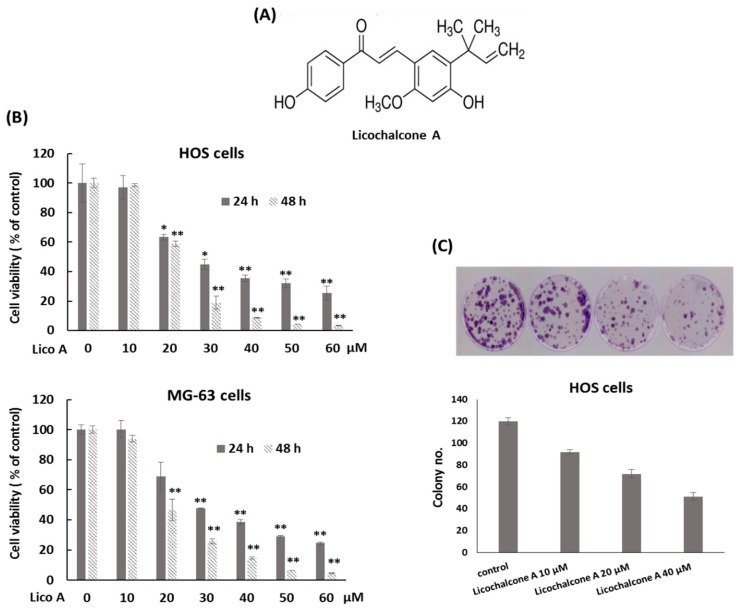
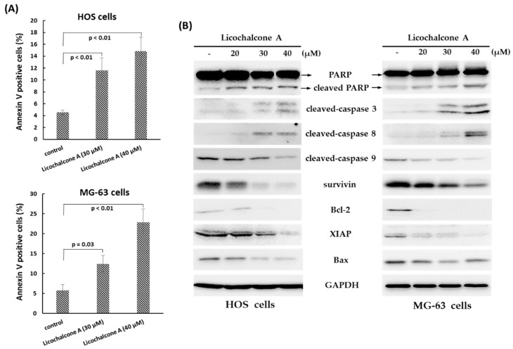
Licochalcone A, a flavonoid extracted from licorice root, has been shown to exhibit broad anti-inflammatory, anti-bacterial, anticancer, and antioxidative bioactivity. In this study, we investigated the antitumor activity of Licochalcone A against human osteosarcoma cell lines. The data showed that Licochalcone A significantly suppressed cell viability in MTT assay and colony formation assay in osteosarcoma cell lines. Exposure to Licochalcone A blocked cell cycle progression at the G2/M transition and induced extrinsic apoptotic pathway in osteosarcoma cell lines. Furthermore, we found the Licochalcone A exposure resulted in rapid ATM and Chk2 activation, and high levels of nuclear foci of phosphorylated Chk2 at Thr 68 site in osteosarcoma cell lines. In addition, Licochalcone A exposure significantly induced autophagy in osteosarcoma cell lines. When Licochalcone A-induced autophagy was blocked by the autophagy inhibitor chloroquine, the expression of activated caspase-3 and Annexin V positive cells were reduced, and cell viability was rescued in Licochalcone A-treated osteosarcoma cell lines. These data indicate that the activation of ATM-Chk2 checkpoint pathway and autophagy may contribute to Licochalcone A-induced anti-proliferating effect in osteosarcoma cell lines. Our findings display the possibility that Licochalcone A may serve as a potential therapeutic agent against osteosarcoma.
Keywords: ATM-Chk2; Licochalcone A; autophagy; osteosarcoma.
Conflict of interest statement
The authors declare no conflict of interest.
Figures


Similar articles
-
A new compound of thiophenylated pyridazinone IMB5043 showing potent antitumor efficacy through ATM-Chk2 pathway.PLoS One. 2018 Feb 2;13(2):e0191984. doi: 10.1371/journal.pone.0191984. eCollection 2018. PLoS One. 2018. PMID: 29394294 Free PMC article.
-
Licochalcone A induces apoptotic cell death via JNK/p38 activation in human nasopharyngeal carcinoma cells.Environ Toxicol. 2019 Jul;34(7):853-860. doi: 10.1002/tox.22753. Epub 2019 Apr 14. Environ Toxicol. 2019. PMID: 30983163
-
Shallot and licorice constituent isoliquiritigenin arrests cell cycle progression and induces apoptosis through the induction of ATM/p53 and initiation of the mitochondrial system in human cervical carcinoma HeLa cells.Mol Nutr Food Res. 2009 Jul;53(7):826-35. doi: 10.1002/mnfr.200800288. Mol Nutr Food Res. 2009. PMID: 19536869
-
Anticancer effects of licochalcones: A review of the mechanisms.Front Pharmacol. 2023 Jan 23;14:1074506. doi: 10.3389/fphar.2023.1074506. eCollection 2023. Front Pharmacol. 2023. PMID: 36755942 Free PMC article.
-
Molecular mechanisms of estrogen receptor β-induced apoptosis and autophagy in tumors: implication for treating osteosarcoma.J Int Med Res. 2019 Oct;47(10):4644-4655. doi: 10.1177/0300060519871373. Epub 2019 Sep 17. J Int Med Res. 2019. PMID: 31526167 Free PMC article. Review.
Cited by
-
Different Cell Responses to Hinokitiol Treatment Result in Senescence or Apoptosis in Human Osteosarcoma Cell Lines.Int J Mol Sci. 2022 Jan 31;23(3):1632. doi: 10.3390/ijms23031632. Int J Mol Sci. 2022. PMID: 35163553 Free PMC article.
-
The COPS3-FOXO3 positive feedback loop regulates autophagy to promote cisplatin resistance in osteosarcoma.Autophagy. 2023 Jun;19(6):1693-1710. doi: 10.1080/15548627.2022.2150003. Epub 2022 Nov 30. Autophagy. 2023. PMID: 36451342 Free PMC article.
-
Oxidative stress, free radicals and antioxidants: potential crosstalk in the pathophysiology of human diseases.Front Chem. 2023 May 10;11:1158198. doi: 10.3389/fchem.2023.1158198. eCollection 2023. Front Chem. 2023. PMID: 37234200 Free PMC article. Review.
-
Perspectives and controversies regarding the use of natural products for the treatment of lung cancer.Cancer Med. 2021 Apr;10(7):2396-2422. doi: 10.1002/cam4.3660. Epub 2021 Mar 2. Cancer Med. 2021. PMID: 33650320 Free PMC article. Review.
-
A ferroptosis-related gene signature associated with immune landscape and therapeutic response in osteosarcoma.Front Oncol. 2022 Nov 11;12:1024915. doi: 10.3389/fonc.2022.1024915. eCollection 2022. Front Oncol. 2022. PMID: 36439512 Free PMC article.
References
-
- Bernthal N.M., Federman N., Eilber F.R., Nelson S.D., Eckardt J.J., Eilber F.C., Tap W.D. Long-Term Results (>25 Years) of a Randomized, Prospective Clinical Trial Evaluating Chemotherapy in Patients with High-Grade, Operable Osteosarcoma. Cancer. 2012;118:5888–5893. doi: 10.1002/cncr.27651. - DOI - PubMed
-
- Kempf-Bielack B., Bielack S.S., Jurgens H., Branscheid D., Berdel W.E., Exner G.U., Gobel U., Helmke K., Jundt G., Kabisch H., et al. Osteosarcoma Relapse after Combined Modality Therapy: An Analysis of Unselected Patients in the Cooperative Osteosarcoma Study Group (Coss) J. Clin. Oncol. 2005;23:559–568. doi: 10.1200/JCO.2005.04.063. - DOI - PubMed
MeSH terms
Substances
Grants and funding
LinkOut - more resources
Full Text Sources
Molecular Biology Databases
Research Materials
Miscellaneous
