

Redox/NIR dual-responsive MoS2 for synergetic chemo-photothermal therapy of cancer
- PMID: 31269964
- PMCID: PMC6607525
- DOI: 10.1186/s12951-019-0510-2
Redox/NIR dual-responsive MoS2 for synergetic chemo-photothermal therapy of cancer
Abstract
Background: The construction of a multifunctional drug delivery system with a variety of advantageous features, including targeted delivery, controlled release and combined therapy, is highly attractive but remains a challenge.
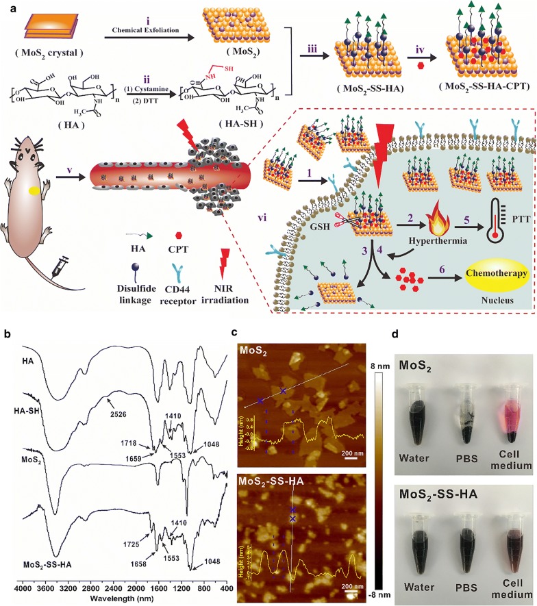
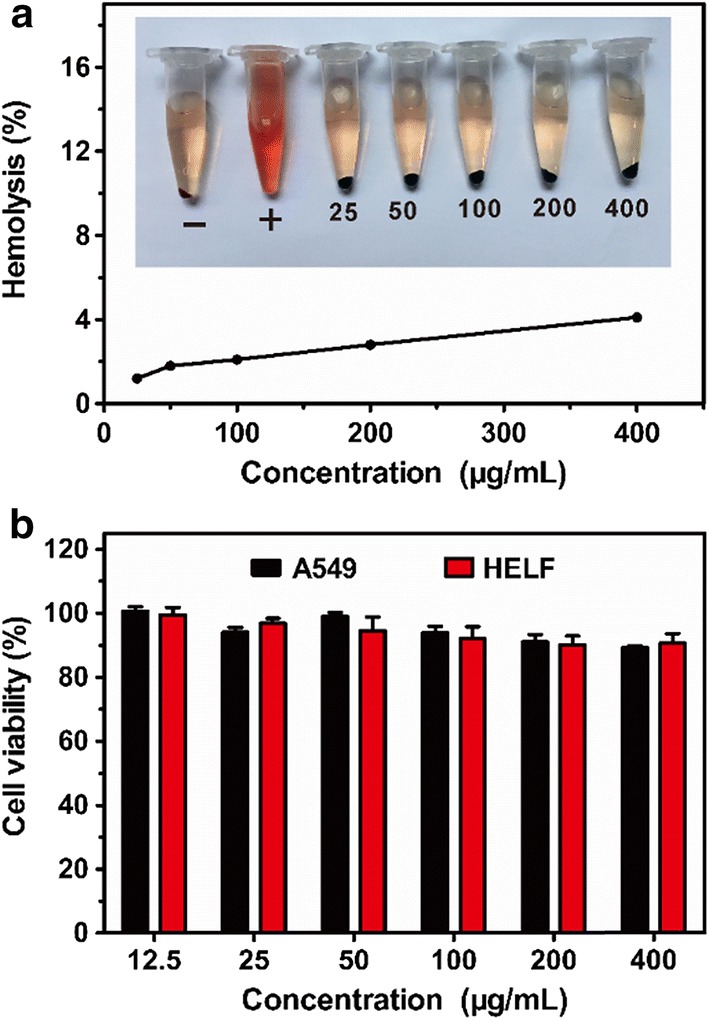
Results: In this study, we developed a MoS2-based hyaluronic acid (HA)-functionalized nanoplatform capable of achieving targeted delivery of camptothecin (CPT) and dual-stimuli-responsive drug release. HA was connected to MoS2 via a disulfide linkage, forming a sheddable HA shell on the surface of MoS2. This unique design not only effectively prevented the encapsulated CPT from randomly leaking during blood circulation but also significantly accelerated the drug release in response to tumor-associated glutathione (GSH). Moreover, the MoS2-based generated heat upon near-infrared (NIR) irradiation could further increase the drug release rate as well as induce photothermal ablation of cancer cells. The results of in vitro and in vivo experiments revealed that MoS2-SS-HA-CPT effectively suppressed cell proliferation and inhibited tumor growth in lung cancer cell-bearing mice under NIR irradiation via synergetic chemo-photothermal therapy.
Conclusions: The as-prepared MoS2-SS-HA-CPT with high targeting ability, dual-stimuli-responsive drug release, and synergistic chemo-photothermal therapy may provide a new strategy for cancer therapy.
Keywords: Chemo-photothermal therapy; Disulfide linkage; Dual-stimuli-responsive drug release; HA; MoS2 nanosheets.
Conflict of interest statement
The authors declare that they have no competing interests.
Figures


Similar articles
-
Molybdenum disulfide-based hyaluronic acid-guided multifunctional theranostic nanoplatform for magnetic resonance imaging and synergetic chemo-photothermal therapy.J Colloid Interface Sci. 2019 Jul 15;548:131-144. doi: 10.1016/j.jcis.2019.04.022. Epub 2019 Apr 8. J Colloid Interface Sci. 2019. PMID: 30991180
-
Functionalized MoS2-erlotinib produces hyperthermia under NIR.J Nanobiotechnology. 2019 Jun 19;17(1):76. doi: 10.1186/s12951-019-0508-9. J Nanobiotechnology. 2019. PMID: 31217009 Free PMC article.
-
A multifunctional nanoplatform based on MoS2-nanosheets for targeted drug delivery and chemo-photothermal therapy.Colloids Surf B Biointerfaces. 2020 Jan 1;185:110585. doi: 10.1016/j.colsurfb.2019.110585. Epub 2019 Oct 17. Colloids Surf B Biointerfaces. 2020. PMID: 31683203
-
An insight into the dual role of MoS2-based nanocarriers in anticancer drug delivery and therapy.Acta Biomater. 2024 Apr 15;179:36-60. doi: 10.1016/j.actbio.2024.03.019. Epub 2024 Mar 27. Acta Biomater. 2024. PMID: 38552760 Review.
-
Recent advances in MoS2-based photothermal therapy for cancer and infectious disease treatment.J Mater Chem B. 2020 Jul 15;8(27):5793-5807. doi: 10.1039/d0tb01018a. J Mater Chem B. 2020. PMID: 32597915 Review.
Cited by
-
Immunostimulatory Potential of MoS2 Nanosheets: Enhancing Dendritic Cell Maturation, Migration and T Cell Elicitation.Int J Nanomedicine. 2020 Apr 29;15:2971-2986. doi: 10.2147/IJN.S243537. eCollection 2020. Int J Nanomedicine. 2020. PMID: 32431496 Free PMC article.
-
Recent developments in two-dimensional molybdenum disulfide-based multimodal cancer theranostics.J Nanobiotechnology. 2024 Aug 28;22(1):515. doi: 10.1186/s12951-024-02785-x. J Nanobiotechnology. 2024. PMID: 39198894 Free PMC article. Review.
-
Multifunctional nanoparticle for cancer therapy.MedComm (2020). 2023 Jan 11;4(1):e187. doi: 10.1002/mco2.187. eCollection 2023 Feb. MedComm (2020). 2023. PMID: 36654533 Free PMC article. Review.
-
Meta-Analysis of Nanoparticle Distribution in Tumors and Major Organs in Tumor-Bearing Mice.ACS Nano. 2023 Oct 24;17(20):19810-19831. doi: 10.1021/acsnano.3c04037. Epub 2023 Oct 9. ACS Nano. 2023. PMID: 37812732 Free PMC article.
-
Intracellular tracking of drug release from pH-sensitive polymeric nanoparticles via FRET for synergistic chemo-photodynamic therapy.J Nanobiotechnology. 2019 Nov 7;17(1):113. doi: 10.1186/s12951-019-0547-2. J Nanobiotechnology. 2019. PMID: 31699100 Free PMC article.
References
MeSH terms
Substances
Grants and funding
LinkOut - more resources
Full Text Sources
Miscellaneous
