

Perplexing case of lung mass perfectly mimicking a malignancy
- PMID: 31270087
- PMCID: PMC6613961
- DOI: 10.1136/bcr-2019-229273
Perplexing case of lung mass perfectly mimicking a malignancy
Abstract
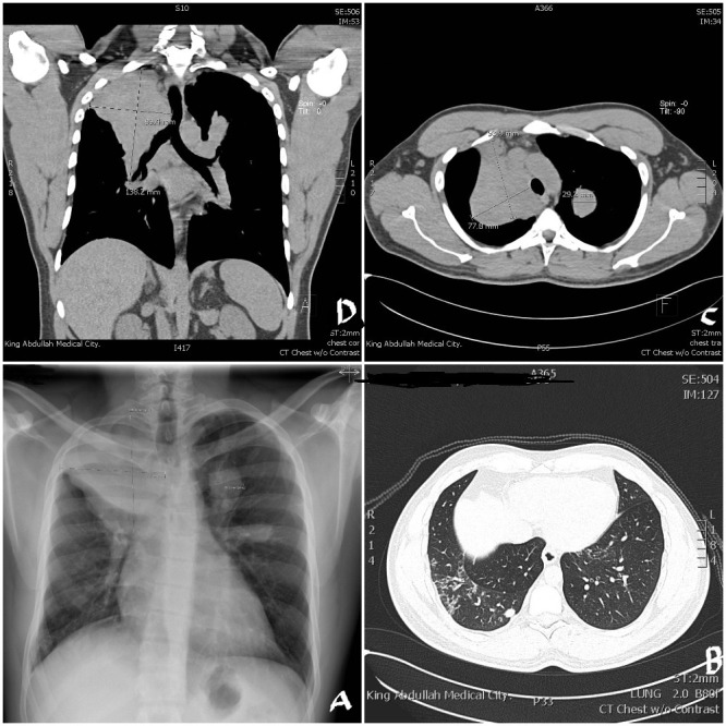
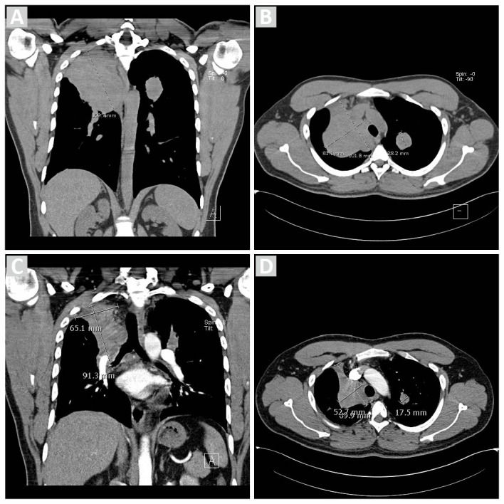
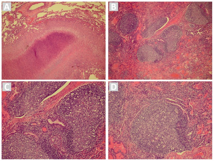
A 35-year-old man, a known asthmatic and with a history of smoking presented with a history of recurrent episodes of mild haemoptysis. On examination, there was decreased intensity of breath sounds on the right infraclavicular area. The chest X-ray and CT chest showed a mass in right upper lobe with nodules in the other lobe. The VAT showed large heavily vascularised mass with surface laden with multiple nodules. The wedge resection of the mass was taken and sent for histopathology examination. The biopsy result showed picture suggestive of connective tissue disease associated follicular bronchiolitis. The patient did not have any signs or symptoms of connective tissue disease. However he was positive for Rheumatoid factor, ANA, anti-RO, anti-CCP antibodies. He was started on steroids and azathioprine. After 6 months of treatment, the size of the mass and nodules reduced by 50% and ESR was reduced to 5 from 75.
Keywords: bronchiolitis; connective tissue disease; interstitial lung disease; rheumatoid arthritis.
© BMJ Publishing Group Limited 2019. No commercial re-use. See rights and permissions. Published by BMJ.
Conflict of interest statement
Competing interests: None declared.
Figures
References
-
- Kang E. Bronchiolitis: Classification and Radiologic Approach. RöFo - Fortschritte auf dem Gebiet der Röntgenstrahlen und der bildgebenden Verfahren [Internet]. Georg Thieme Verlag KG 2006;178(S1).
Publication types
MeSH terms
Substances
LinkOut - more resources
Full Text Sources
Medical
Research Materials
Miscellaneous