. 2019 Aug 15;203(4):873-880.
doi: 10.4049/jimmunol.1900171. Epub 2019 Jul 3.

Identification of the E3 Ligase TRIM29 as a Critical Checkpoint Regulator of NK Cell Functions

Affiliations

Identification of the E3 Ligase TRIM29 as a Critical Checkpoint Regulator of NK Cell Functions

Yaling Dou et al. J Immunol. .

Abstract

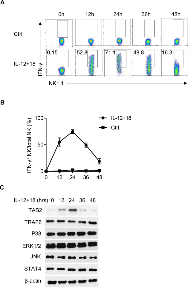
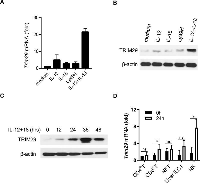
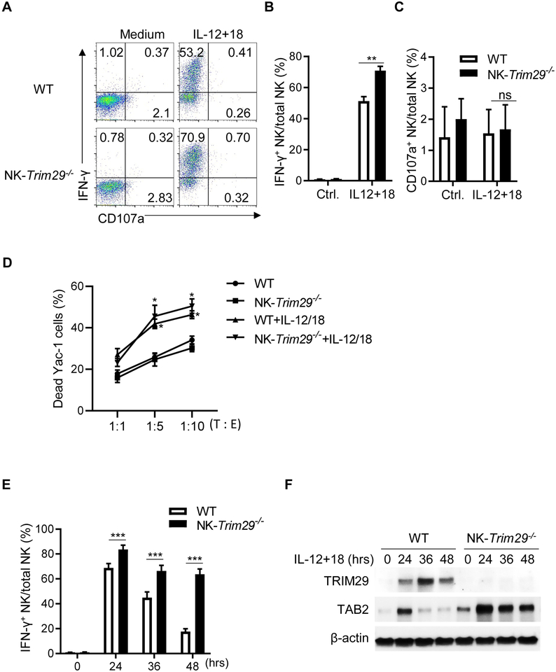
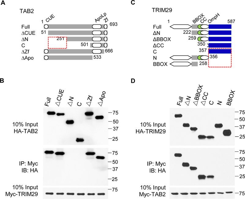
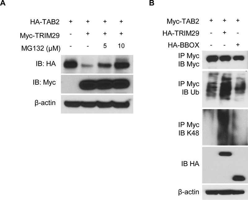
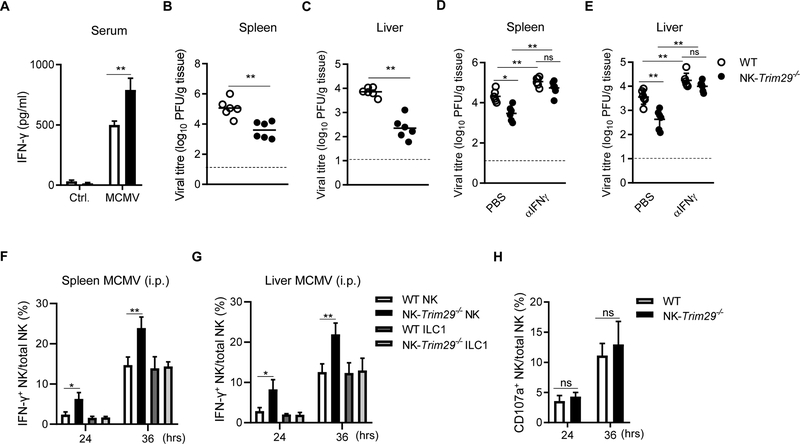
NK cells play an important role in immune surveillance and protective immunity, mainly through rapid cytokine release and cytolytic activities. But how such responses are negatively regulated remains poorly defined. In this study, we demonstrated that the E3 ubiquitin ligase TRIM29 is a crucial regulator of NK cell functions. We found that TRIM29 was not expressed in resting NK cells, but was readily upregulated following activation, especially after IL-12 plus IL-18 stimulation. The levels of TRIM29 expression were inversely correlated with IFN-γ production by NK cells, suggesting that TRIM29 inhibits NK cell functions. Indeed, deficiency of TRIM29, specifically in NK cells, resulted in an enhanced IFN-γ production and consequently protected mice from murine CMV infection. Mechanistically, we showed that once induced in NK cells, TRIM29 ubiquitinates and degrades the TGF-β-activated kinase 1 binding protein 2 (TAB2), a key adaptor protein in IFN-γ production by NK cells. These results identify TRIM29 as a negative regulator of NK cell functions and may have important clinical implications.


Conflict of interest statement

Disclosures

The authors declare no competing financial interests.

Figures

Figure 1.
Figure 1.. The kinetics of IFN-γ expression in NK cells in response to IL-12 and IL-18 stimulation.
(A, B) Splenic NK cells were stimulated with IL-12 (2 ng/ml) and IL-18 (2 ng/ml) in a low concentration of IL-15 (5 ng/ml) for the indicated periods. The expression (A) and the kinetics (B) of IFN-γ were determined by flow cytometry. (C) NK cells were stimulated with IL-12 (2 ng/ml) and IL-18 (2 ng/ml) for 0, 12, 24, 36 or 48 hrs. Cell lysate were prepared for western blot using indicated antibodies. β-actin served as a loading control. Data are representative of 3 independent experiments. Data are mean ± SEM.
Figure 2.
Figure 2.. The induction of TRIM29 in NK cells in response to IL-12 and IL-18 stimulation.
(A) Real-time PCR quantification of Trim29 mRNA in splenic NK cells at 24 hr in response to various combinations of IL-12, IL-18, and plate-bound anti-Ly49H mAbs, shown as fold expression relative to medium control. (B) TRIM29 protein levels were determined by immunoblot at 24 hr. (C) Splenic NK cells were stimulated with IL-12 and IL-18 for the indicated periods and immunoblotted by TRIM29 antibodies. β-actin was used as a loading control. (D) Real-time PCR quantification of Trim29 mRNA in liver CD45+CD3εNK1.1+CD49bCD49a+ ILC1, splenic CD3ε+CD4+ T cells, CD3ε+CD8+ T cells, CD3ε+NK1.1+ NKT cells and CD3εNK1.1+ NK cells in response to IL-12 and IL-18, shown as fold expression relative to naïve CD4+ T cells. Data are representative of 3 independent experiments. Data are mean ± SEM. **P < 0.01; unpaired Student’s t‐test.
Figure 3.
Figure 3.. Sustained TAB2 expression in TRIM29-deleted NK cells promotes IFN-γ production upon cytokines stimulation in vitro.
(A) Flow cytometry plots of IFN-γ and CD107a expression in WT (Trim29f/fNKp46+/+) and NK-Trim29−/− (Trim29f/fNKp46iCre/+) splenic NK cells after 12 hrs upon IL-12 and IL-18 stimulation in vitro. (B and C) Quantification of percentage of intracellular expression of IFN-γ (B) and CD107a production (C) by splenic NK cells by flow cytometric analysis. (D) In vitro cytotoxicity of freshly isolated NK cells or IL-15 (5ng/ml), IL-12 (2 ng/ml) and IL-18 (2 ng/ml)–activated splenic NK cells from WT (Trim29f/fNKp46+/+) and NK-Trim29−/− (Trim29f/fNKp46iCre/+) mice towards YAC-1 targets at the indicated target–to–effector cell ratios (T : E) as quantified by FACS analysis. (E) Quantification of percentage of intracellular expression of IFN-γ by splenic NK cells at 24, 36 and 48 hr after IL-12 and IL-18 stimulation. (F) Immunoblotting of TRIM29 and TAB2 at various time points after stimulation. Data are representative of 3 independent experiments. Data are mean ± SEM. *P < 0.05, **P < 0.01, ***P < 0.001; unpaired Student’s t‐test.
Figure 4.
Figure 4.. Molecular mapping of TRIM29 and TAB2 interactions.
(A) Outline of the constructs of full-length TAB2 and of its truncated mutants: N-terminal coupling of ubiquitin conjugation to ER degradation (CUE), C-terminal zinc finger (ΔZf), and apolipophorin III like (ΔApo). Each construct includes a C-terminal HA tag. (B) Immunoblotting (IB) of purified Myc-tagged TRIM29 with anti-Myc (third blot), purified HA-tagged full-length TAB2 (Full) or its truncations alone (top) or after incubation with Myc-tagged TRIM29 and immunoprecipitation (IP) with anti-Myc (second blot). (C) Outline of the constructs of full-length TRIM29 and of its truncated mutants: the B-box zinc-finger domain (BBOX) and coiled-coil domain (CC). Each construct includes a C-terminal HA tag. (D) Immunoblotting of purified Myc-tagged TAB2 with anti-Myc (third blot), and purified HA-tagged full-length TRIM29 (Full) or its truncations alone (top) or after incubation with Myc-tagged TAB2 and immunoprecipitation with anti-Myc (second blot). Data are representative of 3 independent experiments.
Figure 5.
Figure 5.
TRIM29 induces ubiquitination and degradation of TAB2 via K48 linkage. (A) Immunoblotting of HA-tagged TAB2 with anti-HA (top blot) or of Myc-tagged TRIM29 with anti-Myc (second blot) in HEK293T cells transfected with empty vector or expression vector for Myc-tagged TRIM29 and treated for 4 hrs with 5 or 10 μM of MG132. β-actin was used as a loading control (bottom). (B) Immunoblotting with anti-Myc (top blot), anti-total ubiquitination (ub, 2nd blot), anti-K48-linked ubiquitination (K48, 3rd blot) of Myc-tagged TAB2 in HEK293T cells transfected with empty vector or expression vector for HA-tagged TRIM29, TRIM29-BBOX and treated for 4 hrs with 10 μM of MG132, assessed after immunoprecipitation with anti-Myc, and with anti-HA (4th blot) or anti-β-actin (bottom blot) in whole-cell lysates. Data are representative of 3 independent experiments.
Figure 6.
Figure 6.. TRIM29 deficiency enhances IFN-γ production by NK cells against viral infection.
(A-C) WT (Trim29f/fNKp46+/+) and NK-Trim29−/− (Trim29f/fNKp46iCre/+) mice were infected with MCMV (5 × 10⁴ PFU) intraperitoneally (i.p.). Serum concentration of IFN-γ were measured by ELISA (A), viral titers were measured in spleen (B) and liver (C) at 36 hr post-infection. Dotted line represents the limit of detection for the assay. (D and E) For IFN-γ neutralizing studies, WT and NK-Trim29−/− mice were treated with PBS or α-IFN-γ, followed by infection with MCMV (i.p.). Viral titers were measured in the spleen (D) and liver (E) at 36 hr post-infection. (F and G) Quantification of intracellular of IFN-γ staining of NK cells and ILC1 cells in (F) spleen and (G) liver obtained from WT or NK-Trim29−/− mice following MCMV (i.p.). (H) Quantification of percentage of CD107a in splenic NK cells obtained from WT or NK-Trim29−/− mice following MCMV (i.p.). Data are representative of 3 independent experiments. Data are mean ± SEM. *P < 0.05, **P < 0.01; unpaired Student’s t‐test.

References

    1. Lanier LL 2005. NK cell recognition. Annu Rev Immunol 23: 225–274. - PubMed
    1. Chiossone L, Dumas PY, Vienne M, and Vivier E. 2018. Natural killer cells and other innate lymphoid cells in cancer. Nat Rev Immunol 18: 671–688. - PubMed
    1. Wensveen FM, Jelencic V, Valentic S, Sestan M, Wensveen TT, Theurich S, Glasner A, Mendrila D, Stimac D, Wunderlich FT, Bruning JC, Mandelboim O, and Polic B. 2015. NK cells link obesity-induced adipose stress to inflammation and insulin resistance. Nat Immunol 16: 376–385. - PubMed
    1. Parikh BA, Piersma SJ, Pak-Wittel MA, Yang L, Schreiber RD, and Yokoyama WM. 2015. Dual Requirement of Cytokine and Activation Receptor Triggering for Cytotoxic Control of Murine Cytomegalovirus by NK Cells. PLoS Pathog 11: e1005323. - PMC - PubMed
    1. Gur C, Porgador A, Elboim M, Gazit R, Mizrahi S, Stern-Ginossar N, Achdout H, Ghadially H, Dor Y, Nir T, Doviner V, Hershkovitz O, Mendelson M, Naparstek Y, and Mandelboim O. 2010. The activating receptor NKp46 is essential for the development of type 1 diabetes. Nat Immunol 11: 121–128. - PubMed

Publication types

MeSH terms

Substances