Review
. 2019 Aug 16;294(33):12293-12312.
doi: 10.1074/jbc.REV119.007422. Epub 2019 Jul 2.

Sulfation pathways from red to green

Affiliations
Review

Sulfation pathways from red to green

Süleyman Günal et al. J Biol Chem. .

Abstract

Sulfur is present in the amino acids cysteine and methionine and in a large range of essential coenzymes and cofactors and is therefore essential for all organisms. It is also a constituent of sulfate esters in proteins, carbohydrates, and numerous cellular metabolites. The sulfation and desulfation reactions modifying a variety of different substrates are commonly known as sulfation pathways. Although relatively little is known about the function of most sulfated metabolites, the synthesis of activated sulfate used in sulfation pathways is essential in both animal and plant kingdoms. In humans, mutations in the genes encoding the sulfation pathway enzymes underlie a number of developmental aberrations, and in flies and worms, their loss-of-function is fatal. In plants, a lower capacity for synthesizing activated sulfate for sulfation reactions results in dwarfism, and a complete loss of activated sulfate synthesis is also lethal. Here, we review the similarities and differences in sulfation pathways and associated processes in animals and plants, and we point out how they diverge from bacteria and yeast. We highlight the open questions concerning localization, regulation, and importance of sulfation pathways in both kingdoms and the ways in which findings from these "red" and "green" experimental systems may help reciprocally address questions specific to each of the systems.

Keywords: 3′-phosphoadenosine 5′-phosphosulfate (PAPS) synthase; ATP sulfurylase; Arabidopsis thaliana; adenosine 5′-phosphosulfate kinase; glucosinolates; human; secondary metabolism; steroid hormone; sulfate activation; sulfotransferase; sulfur.


Conflict of interest statement

The authors declare that they have no conflicts of interest with the contents of this article

Figures
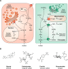
Figure 1.
Figure 1.
Red and green sulfation pathways. A, sulfate is taken up by various sulfate transporters; in plants, some of them transport sulfate into the chloroplast (1). Sulfate activation occurs via animal bifunctional PAPS synthases (2) that shuttle between cytoplasm and nucleus or plant ATP sulfurylase (3) and APS kinase (4) isoforms that are localized in cytoplasm and the chloroplast. PAPS serves as a substrate for cytoplasmic sulfation pathways (5), where PAP is produced. Sulfated compounds can then be de-sulfated by sulfatases (6), enzymes that are absent in plants, or they are secreted via OATPs (7). Two animal PAPS transporters (8) channel PAPS into the Golgi apparatus where many carbohydrate and protein sulfotransferases modify macromolecules for secretion. Although plant protein sulfotransferases are known that reside in the Golgi, an analogous transporter (8) has not yet been identified. Human PAP phosphatases (9) are in the Golgi and the cytoplasm; plant PAP phosphatases are, however, localized in the mitochondrion and the chloroplast. Dedicated PAP(S) transporter in the chloroplast (10) and the mitochondrion (11) deliver PAPS to the cytoplasm and play an important role in the degradation of PAP. In plants, APS represents a branching point where reductive biosynthetic pathways diverge (12). B, examples of structures of sulfated metabolites.
Figure 2.
Figure 2.
PAPS synthases and human sulfotransferases are weakly transcriptionally correlated. Expression profiles for PAPS synthases and sulfotransferases from 27 different human tissues were derived from Fagerberg et al. (48). PAPSS1 and PAPSS2 expression profiles were compared with each other and against different sulfotransferases. Top panel: PAPSS1 and PAPSS2 expression seem to be weakly anti-correlated. Comparing PAPSS1 or PAPSS2 with SULT1A1 shows a weak positive correlation between PAPSS2 and SULT1A1, but a negative correlation for PAPSS1 with SULT1A1. All units in these panels are in RPKM (reads per kb of transcript per million mapped reads). Bottom panel: to illustrate the positive or negative correlation of the tissue-specific expression, the correlation coefficient R is plotted for all 52 sulfotransferases versus PAPSS1 (black) or PAPSS2 (red). There is a tendency for PAPSS2 to be co-expressed with cytosolic sulfotransferases.
Figure 3.
Figure 3.
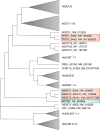
Alignment of sulfotransferases. Protein sequences from 52 human and 21 Arabidopsis sulfotransferases were derived from RefSeq entries from the nucleotide database at www.ncbi.nlm.nih.gov. From multiple splice forms, the one selected was assigned the major isoform. These sequences were subjected to multiple sequence alignments using Clustal Omega and MAFFT (206). From the MAFFT tree, a neighbor-joining tree without distance corrections, a collapsed tree was manually curated. A striking finding was that AtTPST (RefSeq NP_563804) was not grouped with the human TPSTs but with heparan-6-O-sulfotransferases 1–3 (NCBI RefSeq NM_004807, NM_001077188, and NM_153456). The abbreviations used are as follows: SULT, cytosolic sulfotransferase; SOT, plant sulfotransferase; TPST, tyrosylprotein sulfotransferase; CHST, chondroitin sulfotransferase; HS(2/3/6)ST, heparan-(2/3/6)-O-sulfotransferase; NDST, N-deacetylase and N-sulfotransferase; Gal3ST, galactose-3-O-sulfotransferase.
Figure 4.
Figure 4.
Structural representation of different sulfotransferases. A, all structures are shown in the same orientation with the bound PAP nucleotide shown in blue and a substrate in orange. Please note the central β-sheet in all structures and the PAP co-factor are bound exactly at the same position. Structures shown are human sulfotransferase SULT1A1 bound to PAP and 3-cyano-7-hydroxycoumarin (Protein Data Bank code 3U3M), A. thaliana SOT18 complexed to PAP and sinigrin (Protein Data Bank code 5MEX), Danio rerio heparan sulfate 6-O sulfotransferase HS6ST3 with PAP and part of its heptasaccharide displayed (Protein Data Bank code 5T0A), as well as human TPST2 with bound PAP and C4 peptide (Protein Data Bank code 3AP1). Structural visualizations were done using YASARA (207). B, these complexes were structurally aligned using MUSTANG (208). Root mean square deviation (RMSD) values for structural alignment, the number of aligned residues, and the percentage of amino acid identity are listed.

Similar articles

Cited by

References

    1. Beinert H. (2000) A tribute to sulfur. Eur. J. Biochem. 267, 5657–5664 10.1046/j.1432-1327.2000.01637.x - DOI - PubMed
    1. Takahashi H., Kopriva S., Giordano M., Saito K., and Hell R. (2011) Sulfur assimilation in photosynthetic organisms: molecular functions and regulations of transporters and assimilatory enzymes. Annu. Rev. Plant Biol. 62, 157–184 10.1146/annurev-arplant-042110-103921 - DOI - PubMed
    1. Coughtrie M. W. H. (2016) Function and organization of the human cytosolic sulfotransferase (SULT) family. Chem. Biol. Interact. 259, 2–7 10.1016/j.cbi.2016.05.005 - DOI - PubMed
    1. Hirschmann F., Krause F., and Papenbrock J. (2014) The multi-protein family of sulfotransferases in plants: composition, occurrence, substrate specificity, and functions. Front. Plant Sci. 5, 556 10.3389/fpls.2014.00556 - DOI - PMC - PubMed
    1. Mueller J. W., Gilligan L. C., Idkowiak J., Arlt W., and Foster P. A. (2015) The regulation of steroid action by sulfation and desulfation. Endocr. Rev. 36, 526–563 10.1210/er.2015-1036 - DOI - PMC - PubMed

Publication types

LinkOut - more resources