

Newcastle Disease Virus V Protein Degrades Mitochondrial Antiviral Signaling Protein To Inhibit Host Type I Interferon Production via E3 Ubiquitin Ligase RNF5
- PMID: 31270229
- PMCID: PMC6714796
- DOI: 10.1128/JVI.00322-19
Newcastle Disease Virus V Protein Degrades Mitochondrial Antiviral Signaling Protein To Inhibit Host Type I Interferon Production via E3 Ubiquitin Ligase RNF5
Abstract
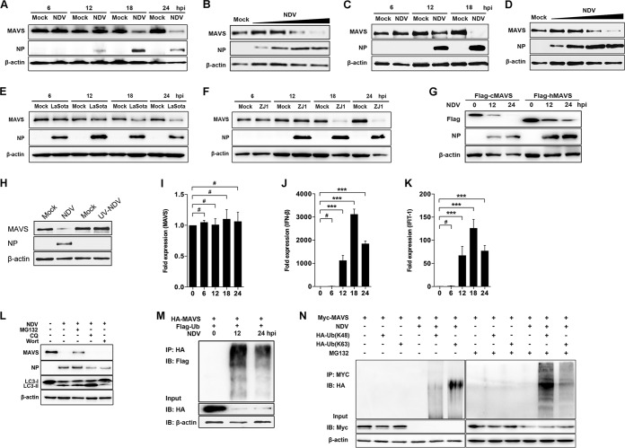
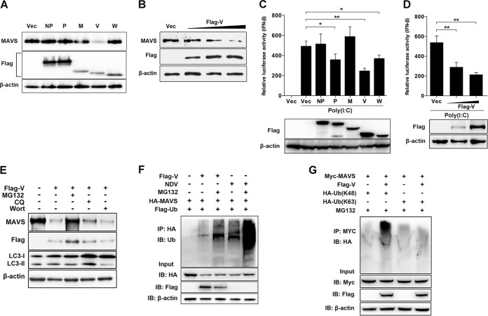
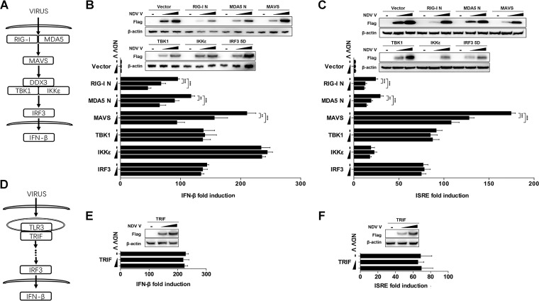
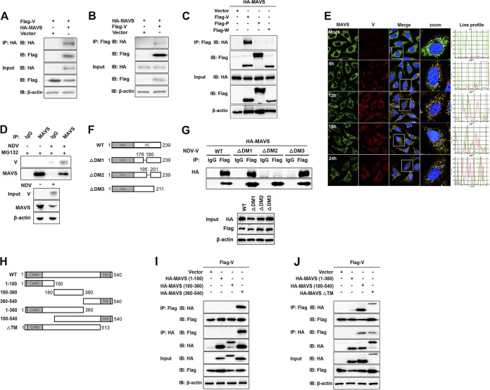
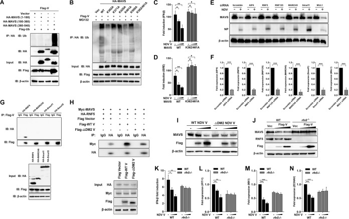
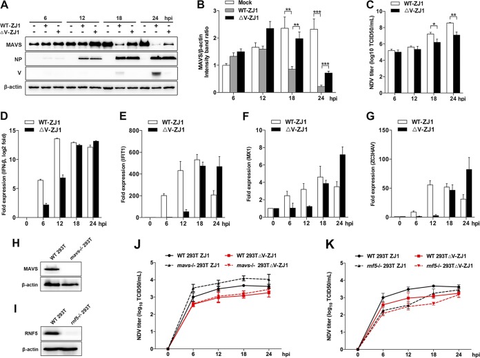
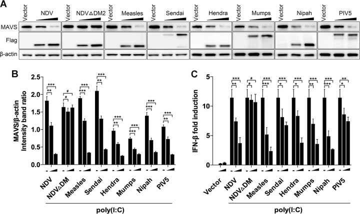
Paramyxovirus establishes an intimate and complex interaction with the host cell to counteract the antiviral responses elicited by the cell. Of the various pattern recognition receptors in the host, the cytosolic RNA helicases interact with viral RNA to activate the mitochondrial antiviral signaling protein (MAVS) and subsequent cellular interferon (IFN) response. On the other hand, viruses explore multiple strategies to resist host immunity. In this study, we found that Newcastle disease virus (NDV) infection induced MAVS degradation. Further analysis showed that NDV V protein degraded MAVS through the ubiquitin-proteasome pathway to inhibit IFN-β production. Moreover, NDV V protein led to proteasomal degradation of MAVS through Lys362 and Lys461 ubiquitin to prevent IFN production. Further studies showed that NDV V protein recruited E3 ubiquitin ligase RNF5 to polyubiquitinate and degrade MAVS. Compared with levels for wild-type NDV infection, V-deficient NDV induced attenuated MAVS degradation and enhanced IFN-β production at the late stage of infection. Several other paramyxovirus V proteins showed activities of degrading MAVS and blocking IFN production similar to those of NDV V protein. The present study revealed a novel role of NDV V protein in targeting MAVS to inhibit cellular IFN production, which reinforces the fact that the virus orchestrates the cellular antiviral response to its own benefit.IMPORTANCE Host anti-RNA virus innate immunity relies mainly on the recognition by retinoic acid-inducible gene I and melanoma differentiation-associated protein 5 and subsequently initiates downstream signaling through interaction with MAVS. On the other hand, viruses have developed various strategies to counteract MAVS-mediated signaling. The mechanism for paramyxoviruses regulating MAVS to benefit their infection remains unknown. In this article, we demonstrate that the V proteins of NDV and several other paramyxoviruses target MAVS for ubiquitin-mediated degradation through E3 ubiquitin ligase RING-finger protein 5 (RNF5). MAVS degradation leads to the inhibition of the downstream IFN-β pathway and therefore benefits virus proliferation. Our study reveals a novel mechanism of NDV evading host innate immunity and provides insight into the therapeutic strategies for the control of paramyxovirus infection.
Keywords: MAVS; NDV; RNF5; degradation; paramyxovirus.
Copyright © 2019 American Society for Microbiology.
Figures
References
-
- Kato H, Takeuchi O, Sato S, Yoneyama M, Yamamoto M, Matsui K, Uematsu S, Jung A, Kawai T, Ishii KJ, Yamaguchi O, Otsu K, Tsujimura T, Koh CS, Reis e Sousa C, Matsuura Y, Fujita T, Akira S. 2006. Differential roles of MDA5 and RIG-I helicases in the recognition of RNA viruses. Nature 441:101–105. doi: 10.1038/nature04734. - DOI - PubMed
Publication types
MeSH terms
Substances
LinkOut - more resources
Full Text Sources
Other Literature Sources
Molecular Biology Databases
Research Materials
Miscellaneous
