Case Reports
. 2019 Sep 2;216(9):2057-2070.
doi: 10.1084/jem.20182295. Epub 2019 Jul 3.

Inherited IFNAR1 deficiency in otherwise healthy patients with adverse reaction to measles and yellow fever live vaccines

Affiliations
Case Reports

Inherited IFNAR1 deficiency in otherwise healthy patients with adverse reaction to measles and yellow fever live vaccines

Nicholas Hernandez et al. J Exp Med. .

Abstract

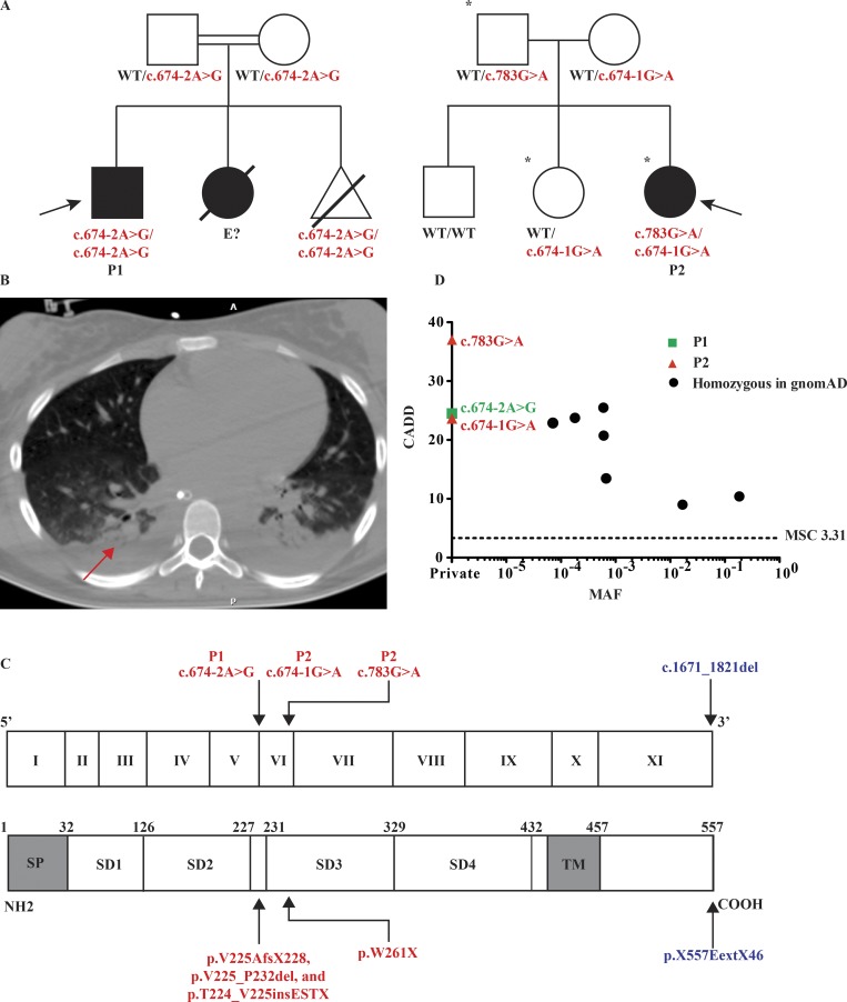
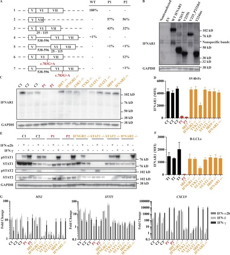
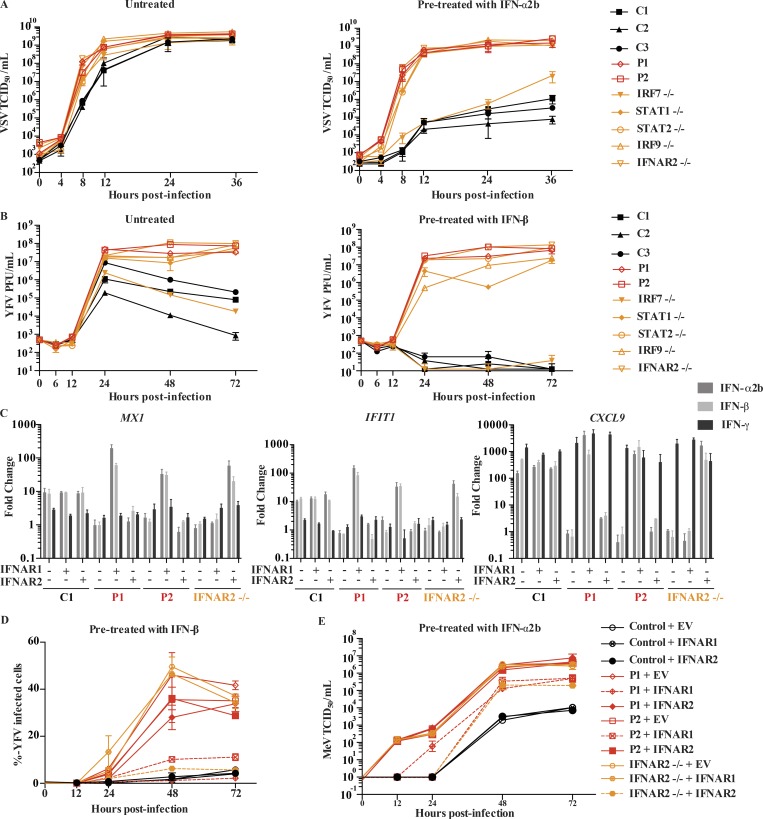
Vaccination against measles, mumps, and rubella (MMR) and yellow fever (YF) with live attenuated viruses can rarely cause life-threatening disease. Severe illness by MMR vaccines can be caused by inborn errors of type I and/or III interferon (IFN) immunity (mutations in IFNAR2, STAT1, or STAT2). Adverse reactions to the YF vaccine have remained unexplained. We report two otherwise healthy patients, a 9-yr-old boy in Iran with severe measles vaccine disease at 1 yr and a 14-yr-old girl in Brazil with viscerotropic disease caused by the YF vaccine at 12 yr. The Iranian patient is homozygous and the Brazilian patient compound heterozygous for loss-of-function IFNAR1 variations. Patient-derived fibroblasts are susceptible to viruses, including the YF and measles virus vaccine strains, in the absence or presence of exogenous type I IFN. The patients' fibroblast phenotypes are rescued with WT IFNAR1 Autosomal recessive, complete IFNAR1 deficiency can result in life-threatening complications of vaccination with live attenuated measles and YF viruses in previously healthy individuals.


Figures

Figure 1.
Figure 1.
Three private IFNAR1 variants are present in two kindreds associated with life-threatening LAV vaccine disease. (A) Pedigrees of the IFNAR1-deficient families. Double lines connecting two parents indicate consanguinity. The probands are indicated by an arrow. Filled shapes indicate affected individuals while open shapes indicate unaffected individuals. Asterisks demarcate those individuals who received vaccination against YFV. The pregnancy of a third sibling in pedigree 1 (homozygous for the same mutation as P1) was terminated at 16 wk of gestational age. (B) CT scan of P2’s chest demonstrating pleural effusions and atelectasis following YF vaccination (red arrow). (C) Schematic illustration of the IFNAR1 gene with 11 coding exons and of the IFNAR1 protein with its four fibronectin type III subdomains (SD1–4). SP, signal peptide; TM, transmembrane domain. The exons are numbered in roman numerals (I–XI). The previously reported IFNAR1 variant is indicated in blue, while those of P1 and P2 are indicated in red. (D) Population genetics of homozygous coding missense and predicted loss-of-function IFNAR1 mutations taken from GnomAD and in-house cohorts. The patients’ variants are private and shown in red and green, while seven variants detected in GnomAD are shown in black. MAF, minor allele frequency; MSC, mutation significance cutoff.
Figure 2.
Figure 2.
Impact of IFNAR1 variants on type I IFN signaling. (A) cDNA sequencing results demonstrate aberrant splicing of IFNAR1 mRNA from patients’ SV40-F cells. The percentage of positively identified transcripts is indicated. At least 150 transcripts were sequenced for each individual. The black numbers under the schematic representations indicate the positions of the included elements relative to their start sites. Results are representative of two experiments. (B) Western blot of IFNAR1 in HEK293T cells transiently transfected with IFNAR1 constructs; GAPDH was used as a loading control. A representative blot from three experiments is shown. (C) Western blot of endogenous IFNAR1 in SV40-F cells from three healthy controls (C1, C2, and C3), P1, P2, IRF7−/−, IFNGR2−/−, TYK2−/−, STAT1−/−, STAT2−/−, IRF9−/−, and IFNAR2−/− patients. A representative blot from three experiments is shown. (D and F) Mean fluorescence intensity (MFI) of IFNAR1 staining on SV40-F (D) and B-LCL (F) cells from three healthy controls (C1, C2, and C3 or T1, T2, and T3), P1, P2, IRF7−/−, IFNGR2−/−, IFNGR1−/−, TYK2−/−, STAT1−/−, STAT2−/−, and IRF9−/− patients as assessed by flow cytometry. Mean (n = 3) and SEM are shown. (E) Western blot analysis of phosphorylated STAT1 (pSTAT1) and pSTAT2 levels in SV40-F cells stimulated with 1,000 U/ml of IFN-α2b or IFN-γ for 20 min. Cells were from two healthy controls (C1, C2), P1, P2, IFNGR2−/−, STAT1−/−, STAT2−/−, and IFNAR2−/− patients. A representative blot from two experiments is shown. (G) Transcription levels of MX1, IFIT1, and CXCL9 assessed by qRT-PCR on SV40-F cells treated with 1,000 U/ml of either IFN-α2b, -β, or -γ for 2 h. Cells were from three healthy controls (C1, C2, and C3), P1, P2, IRF7−/−, IFNGR2−/−, TYK2−/−, STAT1−/−, STAT2−/−, IRF9−/−, and IFNAR2−/− patients. Mean (n = 3) and SEM are shown.
Figure 3.
Figure 3.
IFNAR1 is required for type I IFN–mediated cell intrinsic immunity to viral infections. (A) VSV titers in SV40-F cells unstimulated (left) or pretreated (right) with 1,000 U/ml IFN-α2b for 16 h, followed by VSV infection at an MOI of 3. Cells from three healthy controls were included (C1, C2, and C3), as well as those from P1, P2, IRF7−/−, STAT1−/−, STAT2−/−, IRF9−/−, and IFNAR2−/− patients. Mean (n = 3) and SEM are shown. (B) YFV titers in SV40-F cells unstimulated (left) or pretreated (right) with 1,000 U/ml IFN-β for 16 h, followed by infection with YFV-17D at MOI = 0.05. Mean and range (n = 2) are shown. Cells from three healthy controls were included (C1, C2, and C3), as well as those from P1, P2, IRF7−/−, STAT1−/−, STAT2−/−, IRF9−/−, and IFNAR2−/− patients. (C) Transcription levels of MX1, IFIT1, and CXCL9 assessed by qRT-PCR on SV40-F cells treated with 1,000 U/ml of IFN-α2b, -β, or -γ for 2 h. Cells were from a healthy control, P1, P2, and an IFNAR2−/− patient that were transduced with WT IFNAR1, WT IFNAR2, or EV. Mean (n = 3) and SEM are shown. (D) SV40-F cells were infected with YFV-17D at an MOI of 0.05, and the percentage of cells positive for YFV viral proteins was assessed by flow cytometry. Cells were from a healthy control, P1, P2, and an IFNAR2−/− patient and were transduced with WT IFNAR1, WT IFNAR2, or EV and stimulated with 1,000 U/ml IFN-β for 16 h before infection. Mean and range (n = 2) are shown. (E) SV40-F cells were infected with MeV at an MOI of 0.1, and viral titer was assessed at the given time points. Cells were from a healthy control, P1, P2, and an IFNAR2−/− patient and were transduced with WT IFNAR1, WT IFNAR2, or EV and stimulated with 1,000 U/ml IFN-α2b for 16 h before infection. Mean and SEM (n = 3) are shown. TCID, tissue culture infectious dose.

References

    1. Adzhubei I.A., Schmidt S., Peshkin L., Ramensky V.E., Gerasimova A., Bork P., Kondrashov A.S., and Sunyaev S.R.. 2010. A method and server for predicting damaging missense mutations. Nat. Methods. 7:248–249. 10.1038/nmeth0410-248 - DOI - PMC - PubMed
    1. Belkadi A., Pedergnana V., Cobat A., Itan Y., Vincent Q.B., Abhyankar A., Shang L., El Baghdadi J., Bousfiha A., Alcais A., et al. . Exome/Array Consortium . 2016. Whole-exome sequencing to analyze population structure, parental inbreeding, and familial linkage. Proc. Natl. Acad. Sci. USA. 113:6713–6718. 10.1073/pnas.1606460113 - DOI - PMC - PubMed
    1. Blight K.J., McKeating J.A., and Rice C.M.. 2002. Highly permissive cell lines for subgenomic and genomic hepatitis C virus RNA replication. J. Virol. 76:13001–13014. 10.1128/JVI.76.24.13001-13014.2002 - DOI - PMC - PubMed
    1. Boisson-Dupuis S., Kong X.-F., Okada S., Cypowyj S., Puel A., Abel L., and Casanova J.-L.. 2012. Inborn errors of human STAT1: allelic heterogeneity governs the diversity of immunological and infectious phenotypes. Curr. Opin. Immunol. 24:364–378. 10.1016/j.coi.2012.04.011 - DOI - PMC - PubMed
    1. Bredenbeek P.J., Kooi E.A., Lindenbach B., Huijkman N., Rice C.M., and Spaan W.J.M.. 2003. A stable full-length yellow fever virus cDNA clone and the role of conserved RNA elements in flavivirus replication. J. Gen. Virol. 84:1261–1268. 10.1099/vir.0.18860-0 - DOI - PubMed

Publication types

MeSH terms