. 2019 Aug 13;39(8):BSR20182226.
doi: 10.1042/BSR20182226. Print 2019 Aug 30.

Down-regulated microRNA-30b-3p inhibits proliferation, invasion and migration of glioma cells via inactivation of the AKT signaling pathway by up-regulating RECK

Affiliations

Down-regulated microRNA-30b-3p inhibits proliferation, invasion and migration of glioma cells via inactivation of the AKT signaling pathway by up-regulating RECK

Yan Jian et al. Biosci Rep. .

Retraction in

Abstract

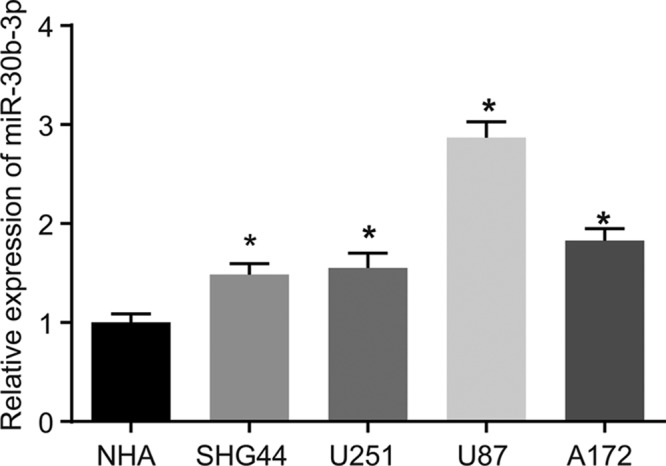
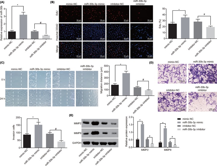
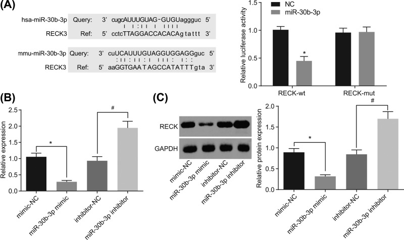
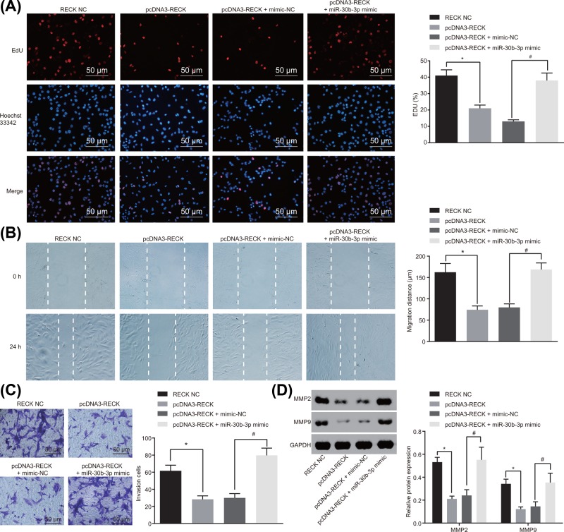
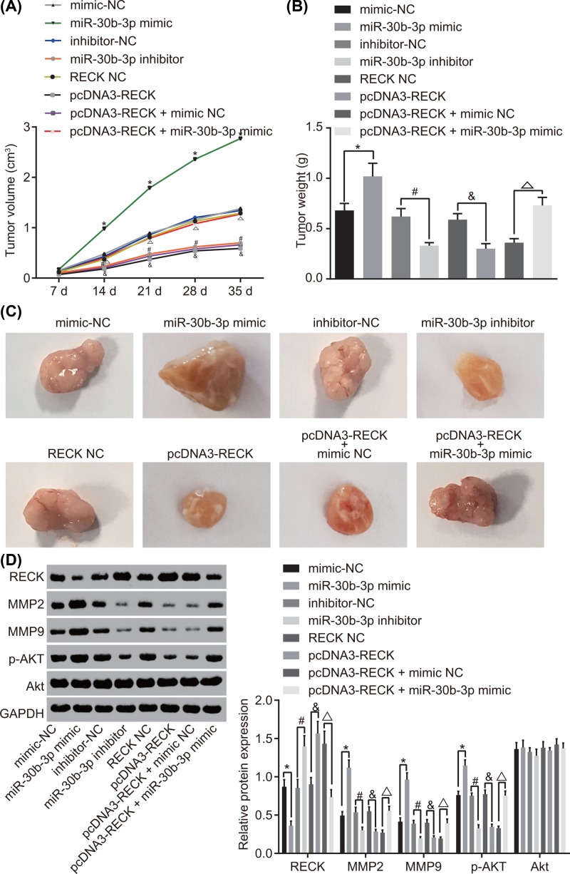
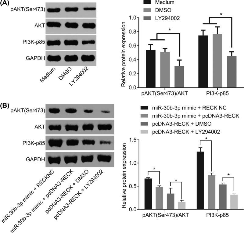
microRNAs (miRNAs) have been found to affect various cancers, and expression of numerous miRNAs is revealed in glioma. However, the role of microRNA-30b-3p (miR-30b-3p) in glioma remains elusive. Therefore, the present study aims to explore the specific mechanism by which miR-30b-3p influence the development of glioma in relation to the AKT signaling pathway. First, glioma cell lines were collected with miR-30b-3p and reversion-inducing cysteine-rich protein with kazal motifs (RECK) expression measured. The functional role of miR-30b-3p and RECK in glioma was determined via gain- and loss-of-function approaches. Subsequently, the expression of invasion- and migration-related factors (MMP-2 and MMP-9) and the AKT signaling pathway-related factors (AKT, p-AKT and PI3K-p85) was detected. Moreover, in vivo experiments were also conducted to investigate how miR-30b-3p influences in vivo tumorigenesis. The results showed that miR-30b-3p was up-regulated and RECK was down-regulated in glioma. RECK was a target gene of miR-30b-3p. Decreased miR-30b-3p and overexpressed RECK led to decreased expression of MMP-2, MMP-9 and p-AKT. Overexpressed RECK and LY294002 could decrease p-AKT and PI3K-p85 expression accompanied with unchanged expression of total protein of AKT. Additionally, proliferation, migration and invasion of glioma cells and tumor formation in nude mice were repressed owing to reduced expression of miR-30b-3p or elevated expression of RECK. In summary, miR-30b-3p inhibition suppresses metastasis of glioma cells by inactivating the AKT signaling pathway via RECK up-regulation, providing a new target for glioma treatment.

Keywords: AKT signaling pathway; Glioma; Invasion; MicroRNA-30b-3p; Migration; Reversion-inducing cysteine-rich protein with kazal motifs.


Conflict of interest statement

The present study was carried out strictly conforming to the recommendations in the Guide for the Care and Use of Laboratory Animals, and was approved by the Animal Ethics Committee of The First Affiliated Hospital of Nanchang University.

The authors declare that there are no competing interests associated with the manuscript.

Figures

Figure 1
Figure 1. miR-30b-3p is overexpressed in glioma cells
*, P<0.05, compared with the NHA cell line; the experiment was repeated three times; data among multiple groups were analyzed by one-way ANOVA and expressed using mean ± SEM; Abbreviation: SEM, standard error of the mean.
Figure 2
Figure 2. Down-regulated miR-30b-3p contributes to inhibited proliferation, migration and invasion of glioma cells
(A) Expression of miR-30b-3p after alteration of miR-30b-3p was detected by RT-qPCR; (B) the viability of glioma cells after alteration of miR-30b-3p was detected by EdU assay (×200); (C) migration ability of glioma cells after alteration of miR-30b-3p was detected by scratch test; (D) invasion ability of glioma cells after alteration of miR-30b-3p was detected by Trasnwell assay (×100); (E) expression of metastasis-associated genes was detected by Western blot analysis; *, P<0.05 compared with the mimic-NC group; #, P<0.05 compared with the inhibitor-NC group; the experiment was repeated three times; the comparison among multiple groups was analyzed by one-way ANOVA, and the data were expressed using mean ± SEM; Abbreviation: SEM, standard error of the mean.
Figure 3
Figure 3. miR-30b-3p targets and down-regulates RECK
(A) Prediction of the binding site between miR-30b-3p and RECK 3′UTR and the target relationship between miR-30b-3p and RECK detected by dual luciferase reporter assay; (B) RECK mRNA level after alteration of miR-30b-3p was detected by RT-qPCR; (C) protein levels of RECK after alteration of miR-30b-3p was detected by Western blot analysis; *, P<0.05 compared with the mimic-NC group; #, P<0.05 compared with the inhibitor-NC group; the experiment was repeated three times; the comparison between two groups was analyzed by one-way ANOVA, and the data were expressed using mean ± SEM; Abbreviation: SEM, standard error of the mean.
Figure 4
Figure 4. U87 cells transfected with pcDNA3-RECK plasmid exhibit overexpression of RECK
(A) Restriction endonuclease digestion of recombinant pcDNA3-RECK plasmid, wherein 1 is DNA Marker, 2 is empty plasmid pcDNA3, 3 and 4 are recombinant plasmid pcDNA3-RECK and 5 is the result of double enzyme digestion of recombinant plasmid pcDNA3-RECK; (B) the expression of RECK in U87 cells transfected with pcDNA3-RECK plasmid was detected by RT-qPCR; *, P<0.05 compared with the RECK NC group; the experiment was repeated three times, and the comparison between groups was analyzed by one-way ANOVA, and the data were expressed using mean ± SEM; Abbreviation: SEM, standard error of the mean.
Figure 5
Figure 5. miR-30b-3p down-regulation suppresses proliferation, migration and invasion of glioma cells by enhancing RECK expression
(A) Viability of glioma cells after alteration of miR-30b-3p and RECK was detected by EdU assay (×200); (B) migration ability of glioma cells after alteration of miR-30b-3p and RECK was detected by scratch test; (C) invasion ability of glioma cells after alteration of miR-30b-3p and RECK was detected by Trasnwell assay (×200); (D) protein levels of metastasis-associated genes after alteration of miR-30b-3p and RECK was detected by Western blot analysis; *, P<0.05 compared with the RECK NC group; #, P<0.05 compared with the pcDNA3-RECK + mimic-NC group; the experiment was repeated three times, and the comparison among multiple groups was analyzed by one-way ANOVA; the data were expressed using mean ± SEM; Abbreviation: SEM, standard error of the mean.
Figure 6
Figure 6. Inhibition of miR-30b-3p repressed tumorigenesis and metastasis in glioma cells in vivo by up-regulating RECK
(A) Tumor volume after alteration of miR-30b-3p and RECK; (B) tumor weight after alteration of miR-30b-3p and RECK on the 35th day; (C) tumor size after alteration of miR-30b-3p and RECK on the 35th day; (D) protein expression of the metastasis-associated genes and the extent of AKT phosphorylation after alteration of miR-30b-3p and RECK detected by Western blot analysis; *, P<0.05 compared with the mimic-NC group; #, P<0.05 compared with the inhibitor-NC group; and P<0.05 compared with the RECK NC group; △, P<0.05 compared with the pcDNA3-RECK + miR-30b-3p NC group; n=12; the comparison among multiple groups was analyzed by one-way ANOVA, and comparison of data at different time points was analyzed using repeated measures analysis of variance; the data were expressed using mean ± SEM; Abbreviation: SEM, standard error of the mean.
Figure 7
Figure 7. miR-30b-3p activates the AKT signaling pathway in glioma cells via RECK down-regulation
(A) Protein expression of AKT signaling pathway-related genes of glioma cells after interference of LY294002 by Western blot analysis. (B) Protein expression of AKT signaling pathway-related genes of glioma cells after interference of miR-30b-3p and RECK by Western blot analysis; *, P<0.05; the experiment was repeated three times; the comparison among multiple groups was analyzed by one-way ANOVA and the data were expressed using mean ± SEM; AKT, Phosphoinositide 3-Kinase/Protein Kinase B; Abbreviation: SEM, standard error of the mean.

Similar articles

Cited by

References

    1. Pereira S., Pinto A., Alves V. and Silva C.A. (2016) Brain tumor segmentation using convolutional neural networks in MRI images. IEEE Trans. Med. Imaging 35, 1240–125110.1109/TMI.2016.2538465 - DOI - PubMed
    1. Omuro A. and DeAngelis L.M. (2013) Glioblastoma and other malignant gliomas: a clinical review. JAMA 310, 1842–185010.1001/jama.2013.280319 - DOI - PubMed
    1. Seliger C., Luber C., Gerken M., Schaertl J., Proescholdt M., Riemenschneider M.J.. et al. (2018) Use of metformin and survival of patients with high-grade glioma. Int. J. Cancer, 144, 273–280 - PubMed
    1. Diamandis P. and Aldape K.D. (2017) Insights from molecular profiling of adult glioma. J. Clin. Oncol. 35, 2386–239310.1200/JCO.2017.73.9516 - DOI - PubMed
    1. Tong B., Pantazopoulou V., Johansson E. and Pietras A. (2018) The p75 neurotrophin receptor enhances HIF-dependent signaling in glioma. Exp. Cell Res. 371, 122–12910.1016/j.yexcr.2018.08.002 - DOI - PubMed

Publication types

MeSH terms