. 2019 Jul 3;11(499):eaau5907.
doi: 10.1126/scitranslmed.aau5907.

The tyrosine kinase inhibitor dasatinib acts as a pharmacologic on/off switch for CAR T cells

Affiliations

The tyrosine kinase inhibitor dasatinib acts as a pharmacologic on/off switch for CAR T cells

Katrin Mestermann et al. Sci Transl Med. .

Abstract

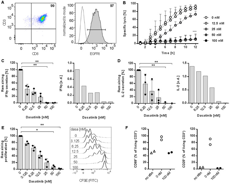
Immunotherapy with chimeric antigen receptor (CAR)-engineered T cells can be effective against advanced malignancies. CAR T cells are "living drugs" that require technologies to enable physicians (and patients) to maintain control over the infused cell product. Here, we demonstrate that the tyrosine kinase inhibitor dasatinib interferes with the lymphocyte-specific protein tyrosine kinase (LCK) and thereby inhibits phosphorylation of CD3ζ and ζ-chain of T cell receptor-associated protein kinase 70 kDa (ZAP70), ablating signaling in CAR constructs containing either CD28_CD3ζ or 4-1BB_CD3ζ activation modules. As a consequence, dasatinib induces a function-off state in CD8+ and CD4+ CAR T cells that is of immediate onset and can be sustained for several days without affecting T cell viability. We show that treatment with dasatinib halts cytolytic activity, cytokine production, and proliferation of CAR T cells in vitro and in vivo. The dose of dasatinib can be titrated to achieve partial or complete inhibition of CAR T cell function. Upon discontinuation of dasatinib, the inhibitory effect is rapidly and completely reversed, and CAR T cells resume their antitumor function. The favorable pharmacodynamic attributes of dasatinib can be exploited to steer the activity of CAR T cells in "function-on-off-on" sequences in real time. In a mouse model of cytokine release syndrome (CRS), we demonstrated that a short treatment course of dasatinib, administered early after CAR T cell infusion, protects a proportion of mice from otherwise fatal CRS. Our data introduce dasatinib as a broadly applicable pharmacologic on/off switch for CAR T cells.


Figures

Fig. 1:
Fig. 1:. Dasatinib locks resting CD8+ CAR-T cells into a function-off state.
(A) Phenotype of CD8+ CAR-T cells. EGFRt is the transduction marker encoded with the CAR-transgene. Numbers indicate percentages of parental population. (B-F) CD8+ CD19-CAR/4-1BB T cells were co-cultured with K562/CD19 in the presence of indicated amounts of dasatinib, which was added at assay start. (B) The percentage of lysed target cells was determined in 1-hour intervals over a period of 12 hours (E:T ratio 5:1). (C, D) ELISA for IFNγ (C) and IL-2 (D). Left diagrams show data obtained in n=3 experiments with T cells from different donors, normalized to the amount of cytokines secreted by CAR-T cells in the absence of dasatinib (100 %). Right diagrams show optical density (o.d.) from one representative experiment. (E) Proliferation of CAR-T cells 72 hours after stimulation with K562/CD19 target cells. Graph shows data from quantitative analysis of n=3 independent experiments. Histograms show data from one representative experiment. The remaining proliferation was calculated using the proliferation index and normalized to the proliferation of CD19-CAR/4-1BB T cells in the absence of dasatinib. (F) Surface expression of activation markers on CAR-T cells without stimulation (no stim) or 20 hours after stimulation with K562/CD19 in the absence (0 nM) and presence (100 nM) of dasatinib. Data shown are mean values + SD (B) or ± SD (C-E) with * P ≤ 0.05, ** P ≤ 0.01 by two-way ANOVA (B) or Kruskal-Wallis test (C-E).
Fig. 2:
Fig. 2:. Dasatinib prevents LCK phosphorylation and NFAT induction after CAR engagement.
For western blot analysis, CD19-CAR/4-1BB T cells were stimulated with CD19+ RCH-ACV and were either treated with 100 nM dasatinib (+) or not (−). (A) Phosphorylation of LCK (Y394), CAR-CD3ζ (Y142), and ZAP70 (Y319) in one representative experiment. (B) Quantitative analysis of western blot data obtained in n=3 experiments, normalized to total protein expression. (C) CD19-CAR/4-1BB T cells were transduced with an NFAT/GFP reporter gene and stimulated with CD19+ Raji or CD19 K562 at an E:T ratio of 5:1. Diagram shows reporter gene expression obtained in CD8+ (left) and CD4+ (right) in the presence (+) or absence (−) of dasatinib (100 nM). (B, C) Data are mean values ± SD analyzed by Kruskal-Wallis test (B) or ordinary one-way ANOVA (C) with * P ≤ 0.05, *** P ≤ 0.001.
Fig. 3:
Fig. 3:. Dasatinib pauses activated CAR-T cells in a function-off state.
CAR-T cells were treated with 100 nM dasatinib added either at assay set-up, or 1, 2, 3, or 48 hours after stimulation with K562/CD19 target cells. (A) Lysis of target cells measured in 1-hour intervals over a 10-hour period. Dasatinib (100 nM) was added 1 hour after assay set-up (orange squares) to switch CAR-T cells off after the initial on phase. For comparison, the diagram shows lysis of target cells by untreated CAR-T cells (blue circles) and by T cells that were treated with dasatinib at assay set-up (red circles). (B) Diagram shows the concentrations of IFNγ (left) and IL-2 (right). Data were normalized to the amount of cytokines produced by untreated CAR-T cells (−, represented by the dotted line). CAR-T cells were treated either at the beginning (0) or 2 hours (2) after assay set-up. (C) Proliferation was calculated based on the cell proliferation index and normalized to proliferation of untreated CAR-T cells (−, represented by the dotted line). (A-C) Data shown are mean values + SD (A) or ± SD (B, C) with * P ≤ 0.05, *** p ≤ 0.001 by two-way ANOVA (A) or Kruskal-Wallis test (B, C).
Fig. 4
Fig. 4. Removal of dasatinib rapidly releases CAR T cells from their function-off state.
(A) Cytolytic activity of CD19-CAR/4-1BB T cells against K562/CD19. Dasatinib (100 nM) was present in the first 2 hours after assay setup (function-off phase) and was removed at t = 0 (function-on phase). Cytolytic activity without dasatinib (0 nM) is shown for comparison. (B to E) T cells were pretreated for 1 or 7 days [dasa pre (+)/1 or 7] with 100 nM dasatinib or kept in culture without dasatinib [dasa pre (−)]. For functional analysis, T cells were washed and cocultured with target cells in the presence [dasa during (+)] or absence [dasa during (−)] of 100 nM dasatinib. (B) Cytolytic capacity of CAR T cells treated with dasatinib at defined schedules. (C and D) Concentrations of IFN-γ (C) and IL-2 (D) after 1 and 7 days of treatment (“Dasa pre”) normalized to the amount of cytokines produced by untreated T cells (−, represented by the dotted line). Dasatinib (100 nM) was added at assay setup in the indicated groups (“Dasa during +”). (E) The proliferation was calculated on the basis of the cell proliferation index and normalized to proliferation of untreated CAR T cells (represented by the dotted line). (F) T cells were treated with 100 nM dasatinib for 7 days (+) or left untreated (−). Viability was analyzed on day 0 (d0; baseline) and on days 2, 4, and 8 by flow cytometry to distinguish alive, early-, and late-stage apoptotic cells (n = 1). (A to E) Data shown are mean values + SD (A) or ± SD (B to E) with **P ≤ 0.01, ***P ≤ 0.001 by two-way (A and B) or one-way ANOVA (C to E). n.s., not significant.
Fig. 5:
Fig. 5:. Dasatinib pauses activated CAR-T cells in a function-off state in vivo.
(A) Treatment schedule and experimental set-up. NSG mice received either 5xl06 CD19-CAR/4-1BB T cells or untransduced T cells (ctrl) on d0 (function-on phase). Dasatinib (10 mg/kg) was administered to indicated groups from d3 to d5 to create a function-off phase. Afterwards, dasatinib was discontinued (function-on phase). (B) Diagram shows average tumor burden (mean) in each treatment group in the function-on-off-on sequence. (C) The ventral BLI signal is displayed for individual mice for up to 94 days after T cell infusion. (D) Bar diagrams show tumor progression/regression in percent during on (d0-3), off (d3-5), and on phase (d5-7) with data for individual mice in each treatment group using CD19-CAR/4-1BB T cells. (E) IFNγ serum concentration during on (d3, d7, d10) and off phases (d5). Data shown are mean and individual values. (F) Bar diagrams show lymphoma progression/regression in percent during the ‘on-off-on’ sequence for individual mice in each treatment group using CD19-CAR/CD28 T cells. (G) The presence of human T cells (CD3+/CD45+) in peripheral blood of mice is depicted as percentage of living cells (7AAD). Blood samples were analyzed by flow cytometry. Shaded areas (B, C, E, and G) indicate the function-off phase. (D-G) Statistical analysis was performed to compare Ctrl (on) and CAR (on) or CAR (on) and CAR (on/off/on) by Mann-Whitney-test (D, F) or two-way ANOVA (E, G) with * P ≤ 0.05, ** P ≤ 0.01, *** P ≤ 0.001.
Fig. 6:
Fig. 6:. Dasatinib mitigates CRS in vivo.
(A) Treatment schedule and experimental set-up. SCID/beige mice received CD19-CAR/CD28 T cells at t=0. Five doses of dasatinib were administered between t=3 and t=33 hours. Afterwards, dasatinib was discontinued. Human (B) and mouse (C) cytokines were measured in serum 8 and 16 hours after T cell infusion. Data shown are mean values from n=3 (ctrl) and n=4 (dasa) treated animals. Statistical analysis was performed using unpaired t-test (B, C) with * P ≤ 0.05, ** P ≤ 0.01, *** P ≤ 0.001. (D) Kaplan-Meyer survival plot for mice receiving CAR and vehicle only (ctrl, n=12) or CAR and dasatinib (dasa, n=13) (statistical analysis by Mantel-Cox test, P=0.0951).

Comment in

References

    1. June CH, Sadelain M, Chimeric Antigen Receptor Therapy, N. Engl. J. Med 379, 64–73 (2018). - PMC - PubMed
    1. Sadelain M, Rivière I, Riddell S, Therapeutic T cell engineering, Nature 545, 423–431 (2017). - PMC - PubMed
    1. Turtle CJ, Hanafi L-A, Berger C, Hudecek M, Pender B, Robinson E, Hawkins R, Chaney C, Cherian S, Chen X, Soma L, Wood B, Li D, Heimfeld S, Riddell SR, Maloney DG, Immunotherapy of non-Hodgkin’s lymphoma with a defined ratio of CD8+ and CD4+ CD19-specific chimeric antigen receptor-modified T cells., Sci. Transl. Med 8, 355ra116 (2016). - PMC - PubMed
    1. Park JH, Rivière I, Gonen M, Wang X, Sénéchal B, Curran KJ, Sauter C, Wang Y, Santomasso B, Mead E, Roshal M, Maslak P, Davila M, Brentjens RJ, Sadelain M, Long-Term Follow-up of CD19 CAR Therapy in Acute Lymphoblastic Leukemia., N. Engl. J. Med 378, 449–459 (2018). - PMC - PubMed
    1. Neelapu SS, Tummala S, Kebriaei P, Wierda W, Gutierrez C, Locke FL, Komanduri KV, Lin Y, Jain N, Daver N, Westin J, Gulbis AM, Loghin ME, de Groot JF, Adkins S, Davis SE, Rezvani K, Hwu P, Shpall EJ, Chimeric antigen receptor T-cell therapy - assessment and management of toxicities., Nat. Rev. Clin. Oncol 15, 47–62 (2018). - PMC - PubMed

Publication types

MeSH terms

Substances