. 2019 Jul 3;9(1):9641.
doi: 10.1038/s41598-019-45997-0.

Conditional loss of ERK1 and ERK2 results in abnormal placentation and delayed parturition in the mouse

Affiliations

Conditional loss of ERK1 and ERK2 results in abnormal placentation and delayed parturition in the mouse

Jessica L Brown et al. Sci Rep. .

Abstract

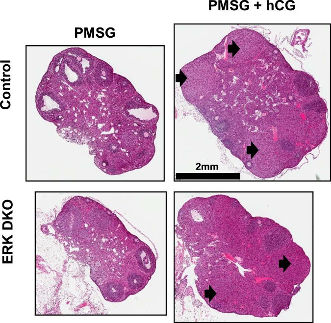
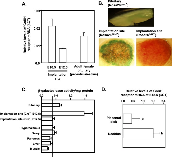
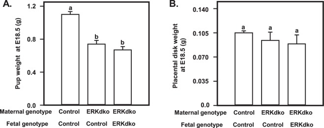
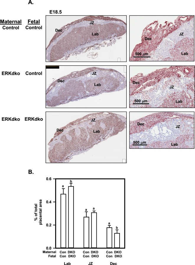
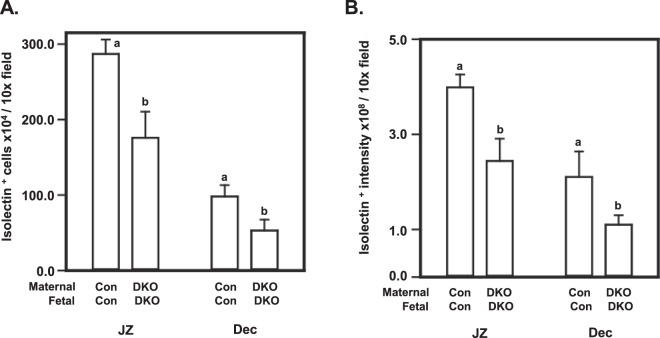
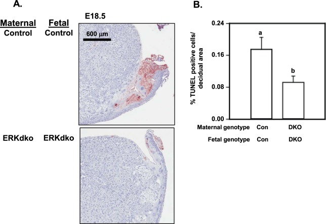
Extracellular-signal-regulated kinases (ERK) 1 and 2 regulate many aspects of the hypothalamic-pituitary-gonadal axis. We sought to understand the role of ERK1/2 signaling in cells expressing a Cre allele regulated by the endogenous GnRHR promoter (GRIC-ERKdko). Adult female GRIC-ERKdko mice were hypogonadotropic and anovulatory. Gonadotropin administration and mating led to pregnancy in one-third of the ERKdko females. Litters from ERKdko females and pup weights were reduced coincident with delayed parturition and 100% neonatal mortality. Based on this, we examined Cre expression in implantation sites as a potential mechanism. GnRHR mRNA levels at e10.5 and e12.5 were comparable to pituitary levels from adult female mice at proestrus and GnRHR mRNA in decidua was enriched compared to whole implantation site. In vivo studies confirmed recombination in decidua, and GRIC-ERKdko placentas showed reduced ERK2 expression. Histopathology revealed abnormalities in placental architecture in the GRIC-ERKdko animals. Regions of apoptosis at the decidual/uterine interface at e18.5 were observed in control animals but apoptotic tone in these regions was reduced in ERKdko animals. These studies support a potential model of ERK-dependent signaling within the implantation site leading to loss of placental architecture and mis-regulation of apoptotic events at parturition occurring coincident with prolonged gestation and neonatal mortality.


Conflict of interest statement

The authors declare no competing interests.

Figures

Figure 1
Figure 1
ERKdko females respond to exogenous gonadotropin stimulation leading to ovulation. Photomicrographs of ovaries from Control and ERKdko female mice following administration of PMSG or PMSG and hCG. Magnification bar is shown and arrows identify some of the corpora lutea.
Figure 2
Figure 2
GnRH receptor (R) mRNA is detectable in the placental disk at embryonic (e) days 10.5 and 12.5 and the GRIC Cre mediates recombination in maternal decidua and placenta. (A) RT-PCR was performed on placental disks from embryonic days 10.5 and 12.5. Levels of GnRHR were comparable with those seen in adult female pituitaries collected at proestrus/estrus. N = 3 placentas/gestational age. In (B) Adult ROSA26GRIC+ mice expressing a β-galactosidase (β-gal)-GFP fusion protein show β-gal activity in the female pituitary in situ. β-gal staining indicates the presence of GRIC Cre activity in placenta from e18.5 ROSA26GRIC+ embryos but not placentas from e18.5 ROSA26GRIC− embryos. (C) Liquid β-gal activity assay confirmed GRIC Cre-mediated recombination in ROSA26GRIC+ pituitaries and placentas. ROSA26GRIC− placental disk and ROSA26GRIC+ hypothalamus, ovary, pancreas, liver and muscle were used as controls. (D) qRT-PCR showed enrichment of GnRHR mRNA expression in placental decidua compared to whole placental disk. Differing letters (a and b) over individual bars identify differences (p < 0.05). N = >4 placentas/genotype.
Figure 3
Figure 3
Pups from GRIC ERKdko females have reduced fetal weight at e18.5. Fetal (A) and placental (B) weights are shown at e18.5 from control/control, GRIC ERKdko/control and GRIC ERKdko/GRIC ERKdko pups and placentas, respectively. N = >4 placentas/genotype. Differing letters (a and b) over individual bars identify differences (p < 0.05).
Figure 4
Figure 4
Histological assessment of placental disks in the GRIC ERKdko model reveals abnormal architecture. (A) Representative sections from control/control, GRIC ERKdko/control and GRIC ERKdko/GRIC ERKdko placentas at e18.5 were stained for isolectin B4 to mark the glycocalyx associated with blood vessel. Panels on the left are low magnification while panels on the right are higher magnification. The decidua (Dec), junctional zone (JZ) and labyrinth (Lab) are indicated. The magnification bar is shown for the higher magnification sections on the right. (B) Quantitation of decidual, junctional zone and labyrinth are reported as % of total placental disk area in control (Con) and GRIC ERKdko (DKO) animals. N = 3 placentas/genotype. Differing letters (a and b) over individual bars identify differences (p < 0.05).
Figure 5
Figure 5
Assessment of isolectin B4 binding in GRIC ERKdko mice implantation sites. The number of isolectin B4+ cells (A) and staining intensity (B) are reported for control/control and GRIC ERKdko dam/GRIC ERKdko groups at e18.5 as number of cells or relative staining intensity (pixel intensity)/10 x magnification field. N = >4 placentas/genotype. Differing letters (a and b) over individual bars identify differences (p < 0.05).
Figure 6
Figure 6
Assessment of TUNEL+ cells within the maternal-fetal interface at near term in GRIC ERKdko females. (A) Representative images of placentas from control/control and GRIC ERKdko/GRIC ERKdko placentas at e18.5 stained using TUNEL as an indicator of apoptosis (red staining). The magnification bar is shown. (B) TUNEL+ cells/μm2 is reported for control/control and GRIC ERKdko/GRIC ERKdko placentas at e18.5. N = 5 placentas/genotype. Differing letters (a and b) over individual bars identify differences (p < 0.05).

Similar articles

Cited by

References

    1. Junttila MR, Li SP, Westermarck J. Phosphatase-mediated crosstalk between MAPK signaling pathways in the regulation of cell survival. FASEB J. 2008;22:954–965. doi: 10.1096/fj.06-7859rev. - DOI - PubMed
    1. Chang L, Karin M. Mammalian MAP kinase signalling cascades. Nature. 2001;410:37–40. doi: 10.1038/35065000. - DOI - PubMed
    1. Bliss SP, et al. ERK signaling in the pituitary is required for female but not male fertility. Mol. Endocrinol. 2009;23:1092–1101. doi: 10.1210/me.2009-0030. - DOI - PMC - PubMed
    1. Bliss SP, Navratil AM, Xie J, Roberson MS. GnRH signaling, the gonadotrope and endocrine control of fertility. Front Neuroendocrinol. 2010;31:322–340. doi: 10.1016/j.yfrne.2010.04.002. - DOI - PMC - PubMed
    1. Shapiro PS, et al. Activation of the MKK/ERK pathway during somatic cell mitosis: direct interactions of active ERK with kinetochores and regulation of the mitotic 3F3/2 phosphoantigen. J. Cell Biol. 1998;142:1533–1545. doi: 10.1083/jcb.142.6.1533. - DOI - PMC - PubMed

Publication types

Substances

LinkOut - more resources