. 2019 Aug;38(31):5987-6001.
doi: 10.1038/s41388-019-0854-y. Epub 2019 Jul 3.

Identification of a novel EphB4 phosphodegron regulated by the autocrine IGFII/IRA axis in malignant mesothelioma

Affiliations

Identification of a novel EphB4 phosphodegron regulated by the autocrine IGFII/IRA axis in malignant mesothelioma

Pierluigi Scalia et al. Oncogene. 2019 Aug.

Erratum in

Abstract

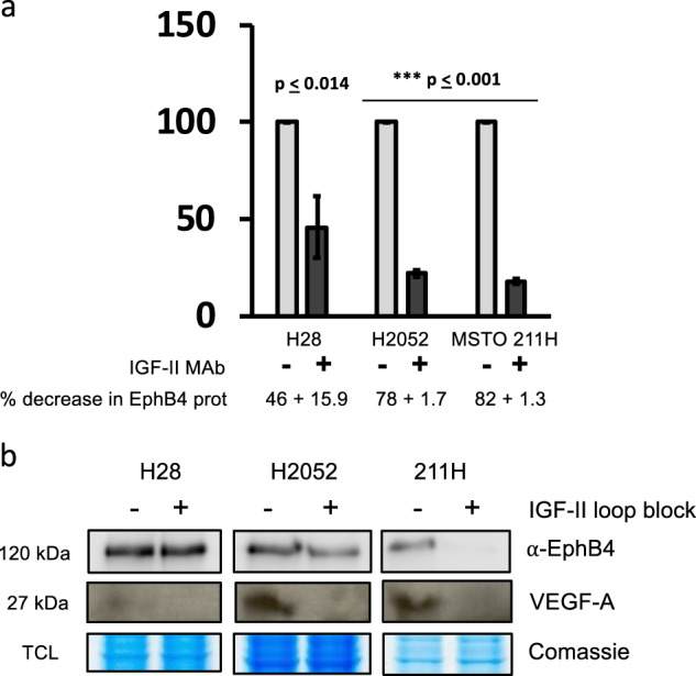
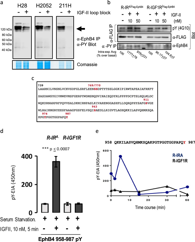
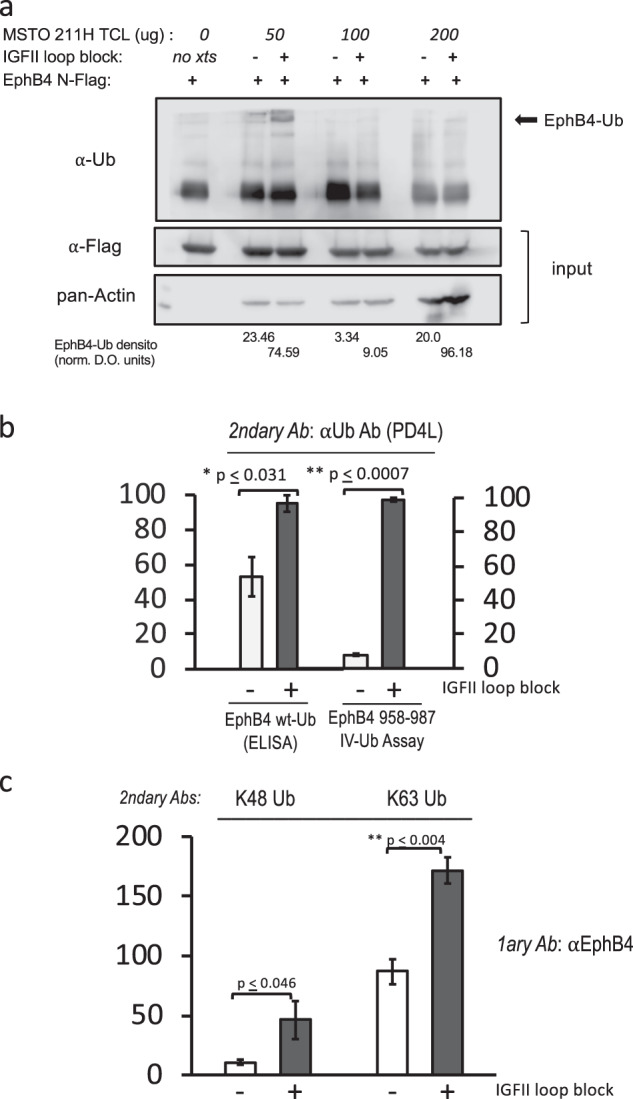
Malignant mesothelioma is a deadly disease with limited therapeutic options. EphB4 is an oncogenic tyrosine kinase receptor expressed in malignant mesothelioma as well as in a variety of cancers. It is involved in tumor microenvironment mediating angiogenesis and invasive cellular effects via both EphrinB2 ligand-dependent and independent mechanisms. The molecular network underlying EphB4 oncogenic effects is still unclear. Here we show that EphB4 expression in malignant mesothelioma cells is markedly decreased upon neutralization of cancer-secreted IGF-II. In particular, we demonstrate that EphB4 protein expression in malignant mesothelioma cells depend upon a degradation rescue mechanism controlled by the autocrine IGF-II-insulin receptor-A specific signaling axis. We show that the regulation of EphB4 expression is linked to a competing post-translational modification of its carboxy-terminal tail via phosphorylation of its tyrosine 987 by the Insulin receptor isoform-A kinase-associated activity in response to the autocrine IGF-II stimuli. Neutralization of this autocrine-induced EphB4-phosphorylation by IGF-II associates with the increased ubiquitination of EphB4 carboxy-terminal tail and with its rapid degradation. We also describe a novel Ubiquitin binding motif in the targeted region as part of the identified EphB4 phosphodegron and provide 3D modeling data supporting a possible model for the acute EphB4 PTM-driven regulation by IGF-II. Altogether, these findings disclose a novel molecular mechanism for the maintenance of EphB4-expression in malignant mesothelioma cells and other IGF-II-secreting cancers (IGF2omas).


Conflict of interest statement

The authors declare that they have no conflict of interest.

Figures

Fig. 1
Fig. 1
IGF-II is found secreted as high molecular weight variants in conditioned media of Mesothelioma cell lines. Secreted IGF-II from conditioned media collected in NCI-H28, NCI-H2052, and NCI-MSTO-211H was detected by immunoprecipitation. The immunocaptured ligand at the end of a neutralization block treatment was collected in prot-A agarose beads, denatured and used for western blotting on a semi-native 15% PAGE. Lanes 1, 4, 7: Genscript markers, lanes 2, 5, 8: conditioned Media control (1/10 input), lanes 3, 6, 9: immunocaptured IGF-II from mesothelioma cell lines conditioned media
Fig. 2
Fig. 2
EphB4 and VEGF-A levels depend upon intact IGF-II autocrine loop in Mesothelioma cell lines. a Column graph of EphB4 levels in mesothelioma cell lines conditionally pre-treated with a neutralizing anti-human IGF-II Ab. EphB4 densitometric values were normalized to the Coomassie staining of TCL and data represented as mean with sem of three experiments. Statistical analysis was performed using two-tailed Student’s t-test. b Immunoblot of EphB4 (upper row), VEGF-A (mid row) and pan-Actin (bottom row). Total cell lysates (TCL, VEGF-A, and pan-Actin) or immunoprecipitates (EphB4) from mesothelioma cancer cell lines conditionally pre-treated with a neutralizing anti-human IGF-II Ab were used
Fig. 3
Fig. 3
The IGF-II autocrine signal is required to insure EphB4 steady-state expression and protein stability in cancer cells. a Reversibility of the antibody-induced autocrine IGF-II block in MSTO 211H. This was evaluated with Ab-induced neutralization of cancer cell-secreted IGF-II followed by release for additional 12 h with fresh conditioned media (without neutralizing Ab) from the same cell line (lanes 2, 4). 50 µg and 200 µg were used for EphB4 immunoprecipitation. b Dependence of the autocrine IGF-II rescue effect on de novo EphB4 synthesized protein by pre-treatment with Cycloheximide for 6 h (left) followed by Ab-neutralization of secreted IGF-II (right). c Time course effect of CHX and IGF-II neutralizing antibody on EphB4 protein levels (without CHX pre-treatment). In each experiment, 15 µl aliquot of the of pre-normalized TCL were used for pan-actin internal control (western blot). The experiments shown are representative of three independent experiments. The experimental workflow used in a, b and c is shown
Fig. 4
Fig. 4
EphB4 is a tyrosine phosphorylative target of IGF-II via activation of the Insulin Receptor isoform A on its Tyr987 residue. a Effect of the autocrine IGF-II signal neutralization on the EphB4 tyrosine phosphorylative status in H28, H2052, and MSTO211H mesothelioma cell lines. Tyrosine phosphorylation was detected by immunoblot in anti-EphB4 immunoprecipitates (upper). The input used for IP was obtained by Coomassie stain (bottom). b R- (MEFs from igf1r null mice) stably expressing either the hIRA or the hIGF1R were transiently transfected with hEphB4 (N-Flag), serum starved for 12 h and stimulated with human synthetic IGF-II at either 10 or 50 nM for 3 min before cell harvesting, solubilization, protein normalization, and anti-Flag (M2) beads or anti-phosphotyrosine immune precipitation. An aliquot (1/10th) of the IP input was used for Flag prot expression control. Intra-experimental average of increased Tyr phosphorylated EphB4 over unstimulated condition is shown. c Known in vivo phosphorylated residues in human EphB4 intracellular region (739–789) sequence obtained from Uniprot (Swiss prot) database showing tyrosine 987 as the only tyrosine residue phosphorylated in vivo in the EphB4 intracellular portion via large phospho-proteomic analysis. d In vitro kinase assay (by EIA) on EphB4 958–987 synthetic substrate using IGF-II pre-stimulated R-hIRA and R-hIGF1R MEF cell lysates after either 5 min stimulation with 10 nM IGF-II post overnight serum starvation. e Time course of human IGF-II stimulation in post-serum starved R-IRA and R-IGF1R MEF cells at 5, 15, 30, and 60 min post-stimulation. The experiments shown are a representative out of three independent experiments. The experiments shown in a are representative of three independent experiments (mean plus SE). Statistical analysis was performed using two-tailed Student’s t-test
Fig. 5
Fig. 5
EphB4 is regulated by ubiquitination and the ubiquitination of its carboxy terminus region is prevented by the IGF-II autocrine stimuli. a In vitro ubiquitination of immobilized N-Flag hEphB4 from transiently expressed R-IRA by MSTO211H cell extracts with or without ex-vivo block of the autocrine IGF-II stimuli (upper row); 1/5th of the immobilized Flag-EphB4 used as substrate for in vitro ubiquitination was used as reaction input control (middle row). The amounts of MSTO211H total cell extracts (50–200 µg) used as source of native ubiquitin ligases in the assay are shown as pan-actin levels (bottom row). The relative rate of ubiquitination based upon densitometric evaluation of the specific bands in the upper row is reported. b Total ubiquitination of endogenous EphB4 immuno-captured from MSTO211H (ELISA, left columns) and the corresponding ubiquitination of immobilized EphB4 958–987 peptide (EIA, right columns) by the same extracts both in presence or absence of autocrine IGF-II block. c Effect of autocrine IGFII block on K48 and K63 Ubiquitin branched proteins in EphB4 immunoprecipitates from MSTO211H. The experiments shown are representative of three independent experiments (mean plus SE for b and c are shown). Statistical analysis for ELISA and EIA experiments in b and c was performed using two-tailed Student’s t-test
Fig. 6
Fig. 6
Identification of a novel Ubiquitin-binding motif on the EphB4 C-terminal region and phylogenetic comparison of C-terminal domain in EphB4. a identification of a combined Ubiquitin-like (green) and a Znf-protein-related (light blue) motif in the EphB4 C-terminus domain (958–974). b Phylogenetic comparison of EphB4 C-Terminus phosphodegron. Green: ubiquitin-like consensus motifs. Red: human sequence conserved residues. Note the C-terminal tyrosine conservation throughout species but in rodents
Fig. 7
Fig. 7
Effect of Tyr987 phosphorylation on EphB4 Carboxy-Terminal 3D conformation and UBX-Znf-Like binding motif access measured as solvent availability by dynamic modeling. Structural consequences of Y987 phosphorylation. a, b Cartoon structure of the SAM domain with the putative UBX-Znf-Like binding domain (amino acids 958 through 974) highlighted in purple. The side chains of the amino acids K973, T976, and Y987 are shown as sticks. The structural models in a, b correspond to the phosphorylated and dephosphorylated state of Y987, respectively. c Solvent accessible surface area (SASA) of the C-terminus loop (amino acids 970 through 987). The SASA was calculated for each frame of the molecular dynamics trajectory; the resulting distributions are shown for the phosphorylated and dephosphorylated cases in cyan and purple, respectively. Note that Y987 phosphorylation causes a statistically significant increase of the SASA

References

    1. Wang HU, Chen ZF, Anderson DJ. Molecular distinction and angiogenic interaction between embryonic arteries and veins revealed by ephrin-B2 and its receptor Eph-B4. Cell. 1998;93:741–53. - PubMed
    1. Erber R, Eichelsbacher U, Powajbo V, Korn T, Djonov V, Lin J, et al. EphB4 controls blood vascular morphogenesis during postnatal angiogenesis. EMBO J. 2006;25:628–41. - PMC - PubMed
    1. Adams RH, Wilkinson GA, Weiss C, Diella F, Gale NW, Deutsch U, et al. Roles of ephrinB ligands and EphB receptors in cardiovascular development: demarcation of arterial/venous domains, vascular morphogenesis, and sprouting angiogenesis. Genes Dev. 1999;13:295–306. - PMC - PubMed
    1. Sinha UK, Kundra A, Scalia P, Smith DL, Parsa B, Masood R, et al. Expression of EphB4 in head and neck squamous cell carcinoma. Ear Nose Throat J. 2003;82:866. - PubMed
    1. Xia G, Kumar SR, Masood R, Zhu S, Reddy R, Krasnoperov V, et al. EphB4 expression and biological significance in prostate cancer. Cancer Res. 2005;65:4623–32. - PubMed

Publication types