Review
. 2019 Jul;14(7):645-657.
doi: 10.1038/s41565-019-0487-x. Epub 2019 Jul 3.

An atlas of nano-enabled neural interfaces

Affiliations
Review

An atlas of nano-enabled neural interfaces

Héctor Acarón Ledesma et al. Nat Nanotechnol. 2019 Jul.

Abstract

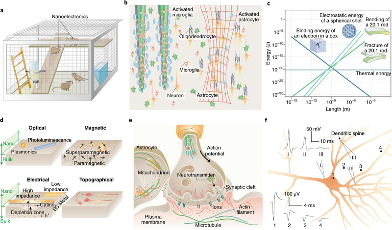
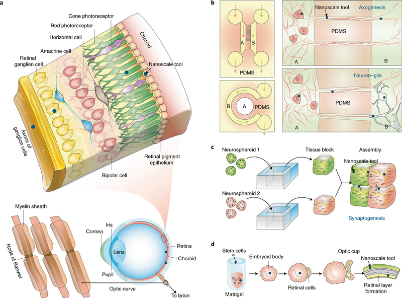
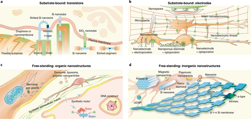
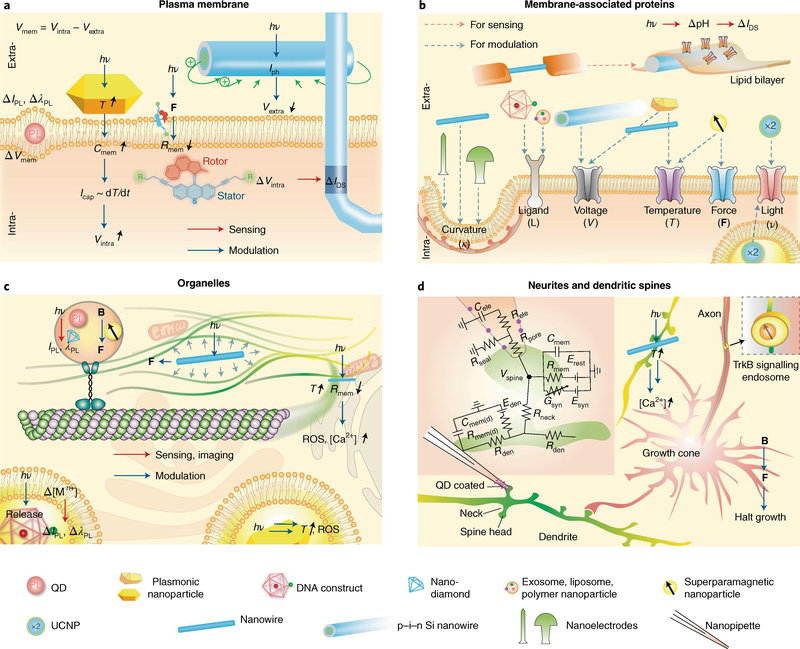
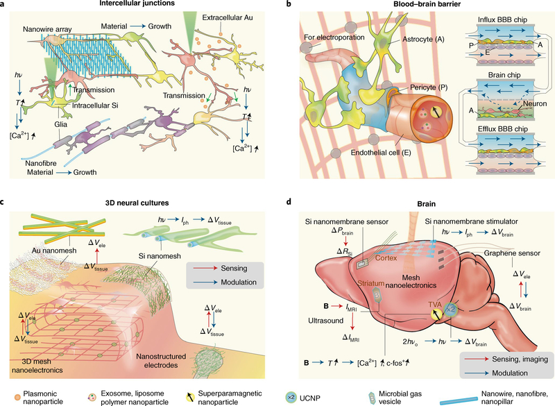
Advances in microscopy and molecular strategies have allowed researchers to gain insight into the intricate organization of the mammalian brain and the roles that neurons play in processing information. Despite vast progress, therapeutic strategies for neurological disorders remain limited, owing to a lack of biomaterials for sensing and modulating neuronal signalling in vivo. Therefore, there is a pressing need for developing material-based tools that can form seamless biointerfaces and interrogate the brain with unprecedented resolution. In this Review, we discuss important considerations in material design and implementation, highlight recent breakthroughs in neural sensing and modulation, and propose future directions in neurotechnology research. Our goal is to create an atlas for nano-enabled neural interfaces and to demonstrate how emerging nanotechnologies can interrogate neural systems spanning multiple biological length scales.


Conflict of interest statement

Competing interests

The authors declare no competing interests.

Figures

Fig. 1 |
Fig. 1 |. Nanoscale materials and devices can offer new opportunities in neural interfaces.
a, Nanodevices offer seamless biointegration with neural components, allowing for imperceptible, minimally invasive and wireless brain interrogation. b, Compared with bulk implants (left), devices with nanoscale thicknesses and open framework are flexible, stretchable and can elicit less immune response (right). Many more astrocytes and microglial cells are recruited and activated at the bulk implant surfaces. c, Many energy terms converge in amplitude at the nanoscale, suggesting that signal transduction at the neural interfaces can take multiple forms,. d, New properties and capabilities emerge at the nanoscale. For example, as the size of metal or semiconductor shrinks, the plasmonic or photoluminescence properties exhibit size- and shape-dependency. Iron oxide switches from paramagnetic to superparamagnetic behaviour when the particle size is smaller than a single magnetic domain. FETs can be gated more easily with nanoscale channel geometries. Nanoscale protrusions or patterns can also yield a neural response that is absent for a planar neural substrate. SC, semiconductor; h+, holes. e, The synaptic junction and many other subcellular domains are highly crowded, dynamic and heterogeneous. Therefore, nanoscale electrophysiology and signal transduction can differ from those recorded in the bulk. f, Electrical recordings show different signal shape and amplitude from different regions of a single neuron. The schematic traces 1–4 are extracellular electrical recordings from points 1–4; traces I–III are intracellular recordings from micropipettes I–III. Adapted from: c, ref. , AIP; ref. , Taylor & Francis.
Fig. 2 |
Fig. 2 |. Naturally occurring and cultured neural systems provide plenty of room for nanoscale probing.
a, Layered structure of retinal tissue. Although epi- and subretinal spaces have been extensively explored for implants, nanoscale tools (blue dots) may access the retinal internal layers. bd, Neural cultures in 2D and 3D can enable bottom-up integration of devices with cells in a seamless manner. b, Microfluidic systems with different compartments (for example, A and B) allow selective manipulation of neural cells, where nanoscale tools (blue dots) can be rationally positioned for studying axogenesis and neuron–glia interactions. c, Neurospheroids can be assembled into 3D tissues, and nanoscale tools (blue dots) can be positioned at the tissue block interfaces for synaptogenesis and circuit studies. d, Embryonic stem cells can develop into lab-grown retina, during which nanoscale tools may be used for growth control or for probing the functions of the final engineered tissues.
Fig. 3 |
Fig. 3 |. Nanoscale toolbox for neural interfaces.
a, Sharp protrusions formed by kinked n–i–n Si nanowires, Si nanotubes, or structures with SiO2 nanotube branches and Si nanowire backbones have enabled transistor-based intracellular electrical recording. Selective etching of a segment in a Si nanowire FET promises localized extracellular recording. 2D nanomaterial-based FETs over flexible substrates can form compliant extracellular biointerfaces. The arrows highlight the entrance of cytosol into FET regions. b, Gold mushroom-shaped nanoelectrodes, nanopillars/nanowires and nanospears yield high-resolution extracellular recording. With electroporation or optoporation, nanopillars/nanowires or nanoporous electrodes achieve intracellular recording. Metal films with nanoscale thicknesses allow brain tissue interfaces with mesh nanoelectronics or nanoelectronic-thread configurations. Nanospears represent an example of integration of a bioelectronics device with a microfluidic system. c, DNA constructs can be used for intracellular delivery and sensing. Synthetic ECM can guide neural differentiation and growth. Exosome, liposome, polymer nanoparticles and synthetic motors may be used for drug delivery to neurons. Recently developed microbial gas vesicles hold promise for MRI and ultrasound imaging. d, Mesoporous Si particles, p–i–n Si nanowires, superparamagnetic nanoparticles, plasmonics nanoparticles, UCNPs and p–i–n Si membranes have been developed for extracellular neuromodulation. The internalized Si nanowires permit intracellular modulation and cytoskeletal control. Quantum dots (QD) and nanodiamonds have been used for imaging or organelle tracking in neurons.
Fig. 4 |
Fig. 4 |. Nano-enabled subcellular neural interfaces.
a, Interfacing plasma membranes: research labs have developed kinked nanowire FETs for intracellular electrical recording, Au nanorods for photothermal neuromodulation, coaxial Si nanowires for photoelectrochemical neuromodulation, synthetic molecular rotors for potential device entrance into neurons, and quantum dots for potential optical recording of membrane voltage. , photon energy; PL, photoluminescence; ΔIPL, PL intensity change; ΔλPL, PL wavelength change; T, temperature; F, force; Icap, capacitive current; Iph, photocurrent; ΔIDS, FET source–drain current change; Cmem, membrane capacitance; Rmem, membrane resistance; Vmem, membrane voltage; Vextra, extracellular potential; Vintra, intracellular potential. b, Interfacing membrane-associated proteins: exosomes, liposomes, polymer nanoparticles and DNA constructs present ligands for cellular targeting and induced endocytosis. Coaxial nanowire produces a photoelectrochemical effect to modulate the local extracellular potential and affects voltage-sensitive ion channels. A rapid photothermal effect from plasmonic nanoparticles or Si nanowires causes depolarization of plasma membrane and triggers activities in voltage-sensitive ion channels. The long-lasting photothermal effect and the magnetothermal effect from superparamagnetic nanoparticles can affect temperature-sensitive ion channels. Superparamagnetic nanoparticles can also actuate mechanical-sensitive ion channels. UCNPs have been used for ion channel modulation, possibly through both extra- and intracellular configuration. Other substrate-bound nanostructures can produce a response from curvature-sensing membrane proteins. Ion channels can also be reconstituted in vitro, producing electrical sensing signals in Si nanowire FET. c, Interfacing organelles: quantum dots or nano-diamonds, plasmonic nanoparticles and superparamagnetic nanoparticles have been used for imaging or controlling cytoskeletal transport or polymerization. Intracellular Au may produce reactive oxygen species (ROS) and other cytotoxic effects upon light illumination. DNA constructs can be used for sensing of calcium or pH in the endosomes. Intracellular Si nanowire also allows for cytoskeletal control via the photoacoustic effect or calcium initiation via the photothermal effect. IPL, PL intensity; λPL, PL wavelength; B, magnetic field; [Mn+], ion concentration. d, Interfacing neuritis and dendritic spines: quantum-dot-coated nanopipette allowed the recording from the spine. Magnetic control of TrkB signalling can halt the neurite growth. A cross-junction between Si nanowire and filapodium produced localized calcium initiation. Inset, schematic diagram of the electrical equivalent circuit for the passive dendritic spine, adapted from ref. . Rden, dendritic resistance; Rele, pipette resistance; Rmem, passive membrane resistance of spine head; Rmem(d), dendrite passive membrane resistance; Rneck, neck resistance; Rpore, pore resistance; Rseal, seal resistance; Cele, pipette capacitance; Cmem, passive membrane capacitance of spine head; Cmem(d), dendrite passive membrane capacitance; Esyn, synaptic reversal potential; Erest, leak reversal potential; Eden, dendritic reversal potential; Gsyn, synaptic conductance. Adapted from: d, ref. , SNL.
Fig. 5 |
Fig. 5 |. Nano-enabled cellular and tissue-scale neural interfaces.
a, Interfacing intercellular junctions. Nanowire arrays can guide neuronal processes and promote intercellular junctions and electrical synchronization. Intracellular Si or extracellular Au-based neural modulations may enable functional connection studies in neurons. Myelination of nano- and microfibres suggests future electrical or optoelectronic control of myelination through semiconductor or metallic nanowires. b, Interfacing the blood–brain barrier. Exosomes, liposomes, polymer nanoparticles and magnetic nanoparticles have been suggested for enhanced delivery through BBB. Future opportunities include localized electroporation through mesh nanoelectronics, given in vitro electroporation studies for enhanced BBB passage. It is also promising to include nanomaterial or nanodevices with organ-on-a-chip model where the interactions among BBB, blood and neural systems can be evaluated together. c, Interfacing 3D neural cultures. Macroporous nanoelectronics mesh can record from the inside of engineered tissues. Conducting nanofibre-coated recording electrodes have low impedance. Au nanomesh used for electrical recording of engineered cardiac tissues and Si nanomesh used for photoelectrochemical modulation of the heart could potentially be used for electrical interfaces with 3D neural cultures. Vele, electrical potential recorded at the nanoelectronics surface; Vtissue, electrical potential from the tissue. d, Interfacing the brain. Biodegradable Si nanomembrane can measure intracranial pressure (Pbrain). Si photodiode nanomembranes produce photoelectrochemical modulation of the cortex and yield a field potential change (ΔVbrain). Semi-transparent graphene electrodes allow flexible electrical recording, and parallel imaging and optical stimulation. A microbial gas vesicle produces MRI signals (IMRI) that can be modulated with ultrasound and allow multiplexed MRI imaging. Superparamagnetic nanoparticles and UCNPs yield deep-brain wireless neuromodulation, producing various effects such as the upregulation of c-fos, a proto-oncogene that is expressed within some neurons following depolarization.

References

    1. Boyden ES, Zhang F, Bamberg E, Nagel G & Deisseroth K Millisecond-timescale, genetically targeted optical control of neural activity. Nat. Neurosci 8, 1263–1268 (2005). - PubMed
    1. Benabid AL Deep brain stimulation for Parkinson’s disease. Curr. Opin. Neurobiol 13, 696–706 (2003). - PubMed
    1. Terem I et al. Revealing sub-voxel motions of brain tissue using phase-based amplified MRI (aMRI). Magn. Reson. Med 80, 2549–2559 (2018). - PMC - PubMed
    1. Salatino JW, Ludwig KA, Kozai TDY & Purcell EK Glial responses to implanted electrodes in the brain. Nat. Biomed. Eng 1, 862–877 (2017). - PMC - PubMed
    1. Insel TR, Landis SC & Collins FS Research priorities. The NIH BRAIN Initiative. Science 340, 687–688 (2013). - PMC - PubMed

Publication types

Substances