. 2019 Jul;25(7):1057-1063.
doi: 10.1038/s41591-019-0498-z. Epub 2019 Jul 3.

Programmable bacteria induce durable tumor regression and systemic antitumor immunity

Affiliations

Programmable bacteria induce durable tumor regression and systemic antitumor immunity

Sreyan Chowdhury et al. Nat Med. 2019 Jul.

Abstract

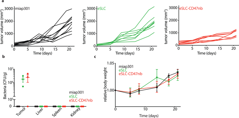
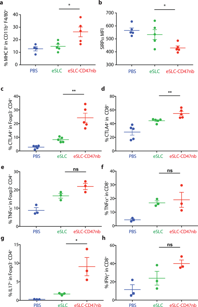
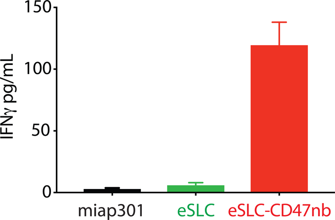
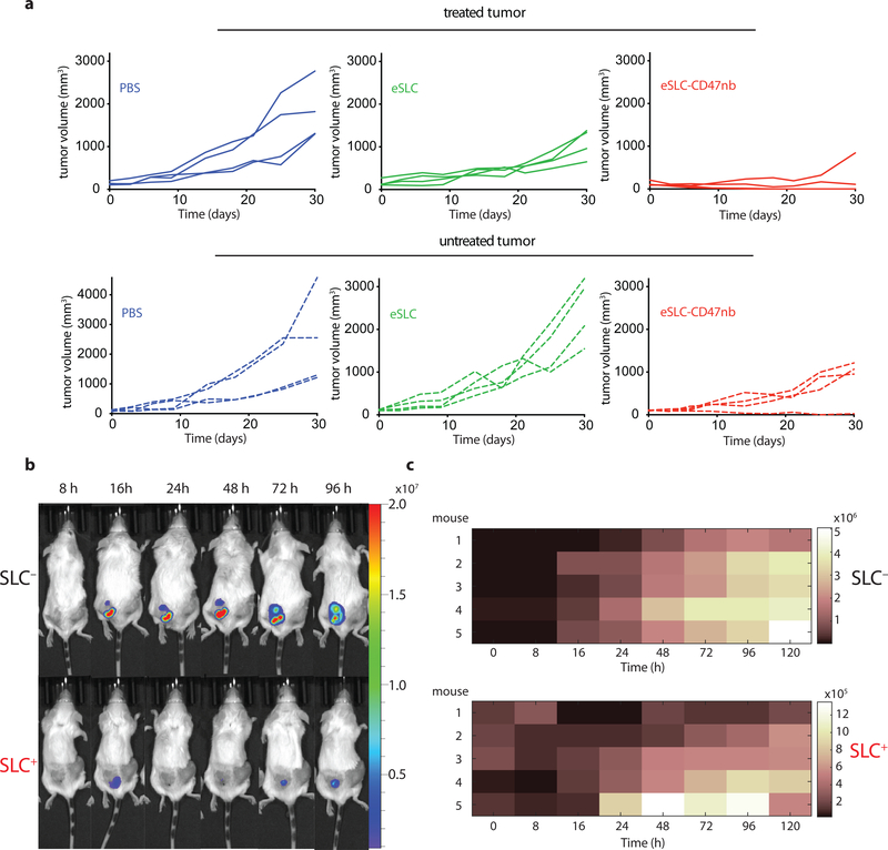
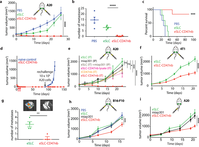
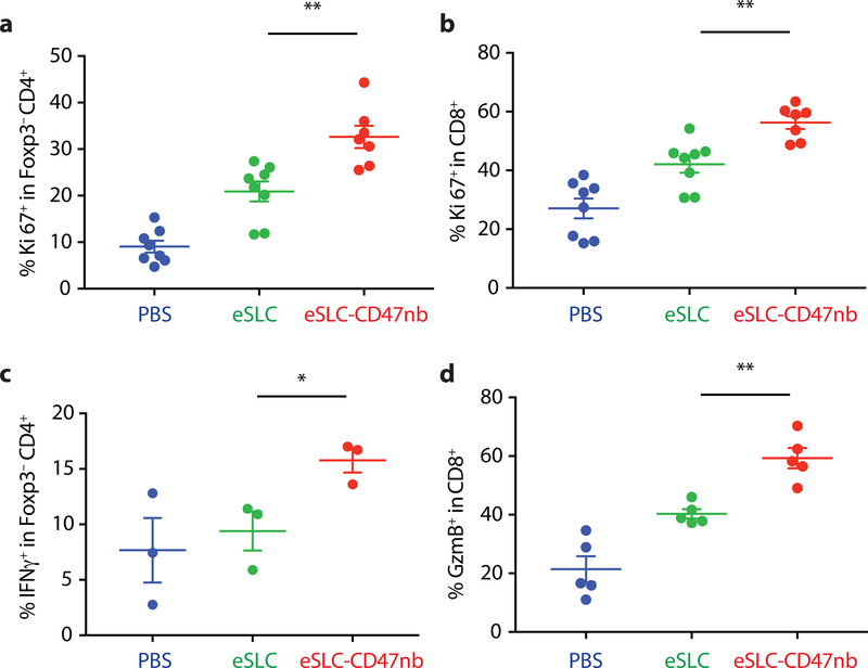
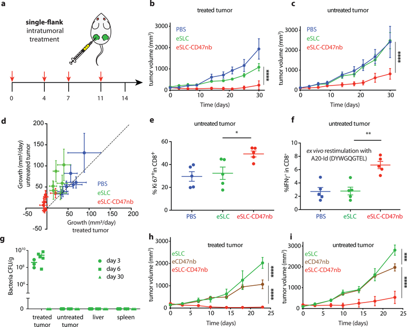
Synthetic biology is driving a new era of medicine through the genetic programming of living cells1,2. This transformative approach allows for the creation of engineered systems that intelligently sense and respond to diverse environments, ultimately adding specificity and efficacy that extends beyond the capabilities of molecular-based therapeutics3-6. One particular area of focus has been the engineering of bacteria as therapeutic delivery systems to selectively release therapeutic payloads in vivo7-11. Here we engineered a non-pathogenic Escherichia coli strain to specifically lyse within the tumor microenvironment and release an encoded nanobody antagonist of CD47 (CD47nb)12, an anti-phagocytic receptor that is commonly overexpressed in several human cancer types13,14. We show that delivery of CD47nb by tumor-colonizing bacteria increases activation of tumor-infiltrating T cells, stimulates rapid tumor regression, prevents metastasis and leads to long-term survival in a syngeneic tumor model in mice. Moreover, we report that local injection of CD47nb-expressing bacteria stimulates systemic tumor-antigen-specific immune responses that reduce the growth of untreated tumors, providing proof-of-concept for an abscopal effect induced by an engineered bacterial immunotherapy. Thus, engineered bacteria may be used for safe and local delivery of immunotherapeutic payloads leading to systemic antitumor immunity.


Conflict of interest statement

COMPETING INTERESTS STATEMENT

S.C., N.A. and T.D. have filed a provisional patent application with the US Patent and Trademark Office (US Patent Application No. 62/747,826) related to this work. T.D. and N.A. have a financial interest in GenCirq, Inc..

Figures

Extended Data Figure 1 |
Extended Data Figure 1 |. Map of plasmids used in this study.
a, pSC01, single plasmid synchronized lysis circuit. b, pSC02, stabilized plasmid driving constitutive expression of HA-tagged anti-CD47 nanobody. c, pSC03 empty vector control.
Extended Data Figure 2 |
Extended Data Figure 2 |. E. coli capable of synchronized lysis produce functional anti-CD47 nanobody.
a, Bacterial growth dynamics over time in agar-pad microscope experiments. b, Immunoblot of bacterial culture supernatants (S) and cell pellets (P) in strains with and without SLC designed to constitutively produce HA-tagged CD47 nanobody. c, A20 cells were co-incubated with a fixed concentration of FITC conjugated IgG2a-FITC isotype control along with varying concentrations of bacterial lysates containing constitutively expressed CD47nb or empty vector. d, A20 cells were co-incubated with a fixed concentration of FITC-conjugated anti-CD47 (miap301) antibody along with serial dilutions of recombinant 6xHis-tagged CD47nb (rCD47nb). e, in vitro phagocytosis of DiI labeled A20 cells pretreated with PBS, miap301, IgG2a isotype control, or serial dilutions of eSLC or eSLC-CD47nb lysate in PBS by bone-marrow derived macrophages.
Extended Data Figure 3 |
Extended Data Figure 3 |. Individual kinetics of intratumoral bacterial immunotherapy.
a, Individual tumor growth trajectories (n=7 per group). b, Representative images of subcutaneous A20 tumor bearing BALB/c mice treated with PBS, eSLC, or eSLC-CD47nb. c, Relative body weight of A20 tumor bearing BALB/c mice over time (ns (not significant), two-way ANOVA with Tukey’s multiple comparisons test). d, Individual tumor growth trajectories (n=4–8 per group) following treatment with eSLC (IT), miap301 (IP), eSLC (IT) + miap301 (IP), eSLC-CD47nb lysate, rCD47nb (IT) or eSLC-CD47nb (IT).
Extended Data Figure 4 |
Extended Data Figure 4 |. Immunotherapeutic bacteria limit tumor growth in syngeneic murine models of melanoma and triple negative breast cancer.
a, Relative body weight of B16-F10 bearing C57BL/6 mice over time (n=4–5 mice per group, ns, two-way ANOVA with Tukey’s multiple comparisons test) b, Individual tumor growth trajectories of subcutaneous 4T1 tumors following intratumoral eSLC or eSLC-CD47nb injection (n=6–8 per group). c, Individual tumor growth trajectories of subcutaneous B16-F10 melanoma following intraperitoneal miap301 or intratumoral PBS, eSLC, or eSLC-CD47nb injection (n=8–12 per group).
Extended Data Figure 5 |
Extended Data Figure 5 |. Intravenous bacterial immunotherapy limits tumor growth in a subcutaneous A20 lymphoma model.
a, Individual tumor growth trajectories of subcutaneous A20 tumors following intraperitoneal miap301 or intravenous eSLC or eSLC-CD47nb treatment (n=8–10 per group). b, Biodistribution of eSLC-CD47nb E. coli on day 8 following final intravenous bacterial treatment. Excised tumors, livers, spleens and kidneys were homogenized, serially diluted and plated on LB agar plates. Colonies were counted to determine CFU/g of tissue (n=3 per group). c, Relative body weight of A20 tumor bearing BALB/c mice receiving intravenous bacterial injections or intraperitoneal injections of miap301 (n=4–5 per group, ns, two-way ANOVA with Tukey’s multiple comparisons test).
Extended Data Figure 6 |
Extended Data Figure 6 |. Immunophenotyping of tumor infiltrating myeloid and lymphoid subsets following intratumoral bacterial injection.
5 × 10⁶ A20 cells were subcutaneously implanted into the hind flanks of BALB/c mice. When tumors reached 100–150 mm3 in volume (day 0), mice were treated with either PBS, eSLC or eSLC-CD47nb on days 0, 4 and 7. On day 3 or day 8, tumors were homogenized and tumor-infiltrating myeloid and lymphoid subsets were isolated for flow cytometric analysis (n=3–5 mice per group) a, Frequency of isolated MHC IIhi CD11b+ F4/80+ macrophages on day 3 following treatment. b, MFI of SIRPα staining within CD11b+ F4/80+ subset on day 8 following treatment. c, d, Frequencies of CTLA4+ within Foxp3CD4+ and CD8+ T cells, respectively. e, f, Frequencies of TNFα+ within Foxp3CD4+ and CD8+ T cells, respectively following ex vivo stimulation. g, Frequency of IL17+ within Foxp3CD4+ T cells following ex vivo stimulation. h, Frequency of IFNγ+ within CD8+ T cells following ex vivo stimulation. (*P<0.05, **P<0.01, unpaired t-test)
Extended Data Figure 7 |
Extended Data Figure 7 |. Immunotherapeutic bacteria lead to increased interferon-γ production by splenic T cells following stimulation with tumor antigens.
IFN-γ ELISA of supernatants from overnight coincubation of splenocytes isolated from each of the indicated treatment groups with irradiated A20 cells (n=2 mice per group, 3 technical replicates).
Extended Data Figure 8 |
Extended Data Figure 8 |. Intratumoral bacterial immunotherapy leads to distal tumor control.
a, Individual tumor growth trajectories of treated (injected) and untreated A20 tumors following intratumoral PBS, eSLC, or eSLC-CD47nb injection. b, SLC and SLC+ EcNisLux were intratumorally injected into a single-flank of A20 tumor bearing mice (scale represents radiance (p/s/cm2/sr). Luminescence was measured over time via IVIS. Representative image of mouse #3 from each group over time. c, Luminescence heat maps over time (n=5 mice per group). Colors represent average radiance (p/s/cm2/sr).
Extended Data Figure 9 |
Extended Data Figure 9 |. Immunophenotyping of tumor infiltrating lymphocytes in untreated tumors following single-flank bacterial injection.
5 × 10⁶ A20 cells were implanted into the hind flanks of BALB/c mice. When tumors reached ~100 mm3 in volume (day 0), mice were treated with either PBS, eSLC or eSLC-CD47nb on day 0, 4 and 7 into a single tumor. Untreated tumors were extracted and analyzed by flow cytometry on day 8. n=5 mice per group. a, Frequency of Ki-67+ cells within Foxp3CD4+ T cells (ns, unpaired t-test). b, c, Frequency of tumor infiltrating IFNγ + within Foxp3CD4+ T cells and CD8+ T cells respectively following ex vivo stimulation with PMA and ionomycin in the presence of brefeldin A (*, P<0.05, unpaired t-test). d, e, Frequencies of CTLA4+ within Foxp3CD4+ T and CD8+ T cells compartments, respectively. (* P<0.05, unpaired t-test). f, Frequency of tumor infiltrating IFNγ + within Foxp3 CD4+ T cells following ex vivo restimulation with A20-Id peptide (DYWGQGTEL) in the presence of brefeldin A (ns, unpaired t-test).
Extended Data Figure 10 |
Extended Data Figure 10 |. Distal tumor control requires SLC+ bacteria engineered to produce CD47nb.
a, b, Individual tumor growth trajectories of treated (injected) and untreated A20 tumors following intratumoral eSLC, eCD47nb, or eSLC-CD47nb injection (n=4 mice per group).
Figure 1 |
Figure 1 |. Quorum-induced release of functional anti-CD47 blocking nanobody by engineered immunotherapeutic bacteria encoding a synchronized lysis circuit (SLC).
a, E. coli with SLC reach a quorum and induce the phage lysis protein ϕX174E, leading to bacterial lysis and release of a constitutively produced, anti-CD47 blocking nanobody which binds to CD47 on the tumor cell surface. b, Bacterial growth dynamics over time of SLC+ and SLC E. coli in batch liquid culture. Data are representative of three independent experimental replicates c, A20 cells were co-incubated with constant concentration of FITC conjugated αCD47 monoclonal antibody (FITC-miap301) along with varying concentrations of bacterial lysates containing constitutively expressed CD47nb (pSC02) or empty vector (pSC03). Data are representative of two independent experimental replicates d, in vitro phagocytosis of DiI labeled A20 cells pretreated with PBS, SLC+ bacteria lysate or SLC+ CD47nb+ bacteria lysate by bone-marrow derived macrophages (n= 4 fields of view × 3 replicates, *** P<0.001, one-way ANOVA with Bonferroni’s multiple comparisons test)
Figure 2 |
Figure 2 |. Intratumoral production of CD47 nanobody by eSLC elicits antitumor responses in multiple syngeneic murine tumor models
a, BALB/c mice (n=7 per group) were implanted subcutaneously with 5 × 10⁶ A20 B-cell lymphoma cells on both hind flanks. When tumor volumes were 100–150 mm3, mice received intratumoral injections every 3–4 days with PBS, eSLC or eSLC-CD47nb in both tumors. Tumor growth curves (**** P<0.0001, two-way ANOVA with Tukey’s multiple comparisons test, error bars represent s.e.m.). Data are representative of two independent experimental replicates b, Quantification of metastatic nodules present in livers on day 30 following bacterial therapy (n= 5 per group, **** P<0.0001, unpaired t-test). c, Kaplan-Meier survival curves for A20 tumor bearing mice (n=5 per group, ***P<0.001, Log-rank (Mantel-Cox test)). d, Mice that had completely cleared A20 tumors were re-challenged with 10 × 106 A20 cells on day 90 following initial treatment. Naive mice received 5 × 10⁶ A20 cells in each flank (n=4 mice per group). e, When A20 tumor volume reached 100–150 mm3 mice received intratumoral injections of eSLC, eSLC-CD47nb bacterial lysate, recombinant CD47nb (rCD47nb, 50 μg) and eSLC-CD47nb, or intraperitoneal injections of anti-CD47 mAb (clone miap-301, 400 μg) alone or in combination with intratumoral eSLC for a total of 4 doses every 3–4 days. Tumor growth curves (n=4–8 per group, **** P<0.0001, *** P<0.001, two-way ANOVA with Tukey’s multiple comparisons test, error bars represent s.e.m.). f, Tumor growth curves. BALB/c mice (n=6–8 per group) were implanted subcutaneously with 106 4T1-Luciferase mammary carcinoma cells. When tumors reached a volume of 200 mm3 mice were randomized and received intratumoral injections of PBS, eSLC, or eSLC-CD47nb every 3 days for a total of 4 doses (**** P<0.0001, two-way ANOVA with Tukey’s multiple comparisons test, error bars represent s.e.m.). Data are representative of two independent experimental replicates. g, IVIS images of lungs extracted from 4T1-Luciferase hind-flank tumors and quantification of number of 4T1-Luciferase metastatic foci in lungs of mice treated with PBS, eSLC or eSLC-CD47nb (** P<0.01, unpaired t-test). h, Tumor growth curves from C57BL/6 mice subcutaneously injected with 5 × 10⁵ B16-F10 melanoma cells into the hind flank. When tumors reached a volume of ~50–150 mm3 mice were randomized and received intraperitoneal injections of miap301 (400 μg) or intratumoral injections of PBS, eSLC, or eSLC-CD47nb every 3 days for a total of 4 doses. (n=8–12 per group. *** P<0.001, two-way ANOVA with Tukey’s multiple comparisons test, error bars represent s.e.m.). i, BALB/c mice were injected with 5 × 10⁶ A20 cells into both hind flanks. When tumor volume reached 100–200 mm3 mice received intravenous injections of eSLC or eSLC-CD47nb or intraperitoneal injections of miap301 CD47mAb (400μg), (n=8–10 per group, **** P<0.0001 twoway ANOVA with Tukey’s multiple comparisons test).
Figure 3 |
Figure 3 |. Immunotherapeutic eSLC-CD47nb bacteria prime robust adaptive antitumor immune responses.
5 × 10⁶ A20 cells were implanted into the hind flanks of BALB/c mice. When tumors reached 100–150 mm3 in volume (day 0), mice were treated with either PBS, eSLC or eSLC-CD47nb on days 0, 4 and 7. On day 8 tumors were homogenized and tumor-infiltrating lymphocytes were isolated for flow cytometric analysis on day 8. a, b Frequencies of isolated intratumoral Ki-67+ Foxp3CD4+ and CD8+ T cells. c, Tumor infiltrating lymphocytes were stimulated following ex vivo isolation with PMA and ionomycin in the presence of brefeldin A. Frequencies of intratumoral IFNγ+ Foxp3CD4+ T cells following stimulation. d, Percentages of intratumoral Granzyme-B positive CD8+ T cells. (n= 3–7 per group. * P<0.05, ** P<0.01, unpaired t-test, error bars represent s.e.m.). Data are pooled from two independent experimental replicates.
Figure 4 |
Figure 4 |. Systemic adaptive immunity following bacterial therapy limits growth of untreated tumors.
a, Treatment schedule. BALB/c mice (n=4 per group) were implanted subcutaneously with 5 × 10⁶ A20 cells on both hind flanks. When tumor volumes reached 100–150 mm3, mice received intratumoral injections every 3–4 days with PBS, eSLC or eSLC-CD47 into a single tumor. b, c Tumor growth curves of treated and untreated tumors (**** P<0.0001, two-way ANOVA with Tukey’s multiple comparisons test, error bars represent s.e.m.). Data are representative of three independent experimental replicates d, Plot of untreated tumor growth rate (mm3/day) vs. treated tumor growth rate (mm3/day) for each mouse. Dotted line indicates slope=1, points represent means, error bars represent s.e.m. e, Untreated tumors were isolated on day 8 following single flank bacterial injections and analyzed by flow cytometry. Frequencies of intratumoral Ki-67+ CD8+ T cells (n=5 per group, * P<0.05, unpaired t-test, error bars represent s.e.m.). Data are representative of two independent experimental replicates. f, Tumor infiltrating lymphocytes were stimulated following ex vivo isolation with A20-Id peptide (DYWGQGTEL) in the presence of brefeldin A. Frequencies of intratumoral IFNγ+ CD8+ T cells (n=5 per group, ** P<0.01, unpaired t-test, error bars represent s.e.m.) g, Biodistribution of SLC+ E. coli on day 3, 6 and 30 following intratumoral bacterial injection. Excised tumors, livers and spleens were homogenized and plated on LB agar plates. Colonies were counted to determine CFU/g of tissue. Limit of detection 103 CFU/g (n=3–5 per time point). h, i Tumor growth curves of treated and untreated A20 tumors following unilateral intratumoral injections of eSLC, eCD47nb or eSLC-CD47nb every 3–4 days for a total of 4 doses (n=4 per group, *** P<0.001, **** P<0.0001, two-way ANOVA with Tukey’s multiple comparisons test, error bars represent s.e.m.).

Comment in

Similar articles

Cited by

References

    1. Fischbach MA, Bluestone JA & Lim WA Cell-based therapeutics: the next pillar of medicine. Sci Transl Med 5, 179ps177 (2013). - PMC - PubMed
    1. Weber W & Fussenegger M Emerging biomedical applications of synthetic biology. Nat Rev Genet 13, 21–35 (2011). - PMC - PubMed
    1. Lim WA & June CH The Principles of Engineering Immune Cells to Treat Cancer. Cell 168, 724–740 (2017). - PMC - PubMed
    1. Ruder WC, Lu T & Collins JJ Synthetic biology moving into the clinic. Science 333, 1248–1252 (2011). - PubMed
    1. Chen YY & Smolke CD From DNA to targeted therapeutics: bringing synthetic biology to the clinic. Sci Transl Med 3, 106ps142 (2011). - PMC - PubMed

Publication types