Skeletal Mineralization in Association with Type X Collagen Expression Is an Ancestral Feature for Jawed Vertebrates
- PMID: 31270539
- PMCID: PMC6759074
- DOI: 10.1093/molbev/msz145
Skeletal Mineralization in Association with Type X Collagen Expression Is an Ancestral Feature for Jawed Vertebrates
Abstract
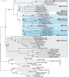
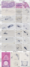
In order to characterize the molecular bases of mineralizing cell evolution, we targeted type X collagen, a nonfibrillar network forming collagen encoded by the Col10a1 gene. It is involved in the process of endochondral ossification in ray-finned fishes and tetrapods (Osteichthyes), but until now unknown in cartilaginous fishes (Chondrichthyes). We show that holocephalans and elasmobranchs have respectively five and six tandemly duplicated Col10a1 gene copies that display conserved genomic synteny with osteichthyan Col10a1 genes. All Col10a1 genes in the catshark Scyliorhinus canicula are expressed in ameloblasts and/or odontoblasts of teeth and scales, during the stages of extracellular matrix protein secretion and mineralization. Only one duplicate is expressed in the endoskeletal (vertebral) mineralizing tissues. We also show that the expression of type X collagen is present in teeth of two osteichthyans, the zebrafish Danio rerio and the western clawed frog Xenopus tropicalis, indicating an ancestral jawed vertebrate involvement of type X collagen in odontode formation. Our findings push the origin of Col10a1 gene prior to the divergence of osteichthyans and chondrichthyans, and demonstrate its ancestral association with mineralization of both the odontode skeleton and the endoskeleton.
Keywords: chondrichthyan; mineralization; scales; teeth; type X collagen.
© The Author(s) 2019. Published by Oxford University Press on behalf of the Society for Molecular Biology and Evolution.
Figures


Similar articles
-
Evolution of dental tissue mineralization: an analysis of the jawed vertebrate SPARC and SPARC-L families.BMC Evol Biol. 2018 Aug 30;18(1):127. doi: 10.1186/s12862-018-1241-y. BMC Evol Biol. 2018. PMID: 30165817 Free PMC article.
-
Molecular footprinting of skeletal tissues in the catshark Scyliorhinus canicula and the clawed frog Xenopus tropicalis identifies conserved and derived features of vertebrate calcification.Front Genet. 2015 Sep 15;6:283. doi: 10.3389/fgene.2015.00283. eCollection 2015. Front Genet. 2015. PMID: 26442101 Free PMC article.
-
Parallel Evolution of Ameloblastic scpp Genes in Bony and Cartilaginous Vertebrates.Mol Biol Evol. 2022 May 3;39(5):msac099. doi: 10.1093/molbev/msac099. Mol Biol Evol. 2022. PMID: 35535508 Free PMC article.
-
Role of elasmobranchs and holocephalans in understanding peptide evolution in the vertebrates: Lessons learned from gonadotropin releasing hormone (GnRH) and corticotropin releasing factor (CRF) phylogenies.Gen Comp Endocrinol. 2018 Aug 1;264:78-83. doi: 10.1016/j.ygcen.2017.09.013. Epub 2017 Sep 19. Gen Comp Endocrinol. 2018. PMID: 28935583 Review.
-
Mineralized Cartilage and Bone-Like Tissues in Chondrichthyans Offer Potential Insights Into the Evolution and Development of Mineralized Tissues in the Vertebrate Endoskeleton.Front Genet. 2021 Dec 22;12:762042. doi: 10.3389/fgene.2021.762042. eCollection 2021. Front Genet. 2021. PMID: 35003210 Free PMC article. Review.
Cited by
-
Regenerating zebrafish scales express a subset of evolutionary conserved genes involved in human skeletal disease.BMC Biol. 2022 Jan 21;20(1):21. doi: 10.1186/s12915-021-01209-8. BMC Biol. 2022. PMID: 35057801 Free PMC article.
-
Endoskeletal mineralization in chimaera and a comparative guide to tessellated cartilage in chondrichthyan fishes (sharks, rays and chimaera).J R Soc Interface. 2020 Oct;17(171):20200474. doi: 10.1098/rsif.2020.0474. Epub 2020 Oct 14. J R Soc Interface. 2020. PMID: 33050779 Free PMC article.
-
Enzymatic Approach in Calcium Phosphate Biomineralization: A Contribution to Reconcile the Physicochemical with the Physiological View.Int J Mol Sci. 2021 Nov 30;22(23):12957. doi: 10.3390/ijms222312957. Int J Mol Sci. 2021. PMID: 34884758 Free PMC article. Review.
-
Transcriptomic landscape of Atlantic salmon (Salmo salar L.) skin.G3 (Bethesda). 2023 Nov 1;13(11):jkad215. doi: 10.1093/g3journal/jkad215. G3 (Bethesda). 2023. PMID: 37724757 Free PMC article.
-
Collagen X Is Dispensable for Hypertrophic Differentiation and Endochondral Ossification of Human iPSC-Derived Chondrocytes.JBMR Plus. 2023 Mar 29;7(5):e10737. doi: 10.1002/jbm4.10737. eCollection 2023 May. JBMR Plus. 2023. PMID: 37197316 Free PMC article.
References
-
- Aldea D, Hanna P, Munoz D, Espinoza J, Torrejon M, Sachs L, Buisine N, Oulion S, Escriva H, Marcellini S.. 2013. Evolution of the vertebrate bone matrix: an expression analysis of the network forming collagen paralogues in amphibian osteoblasts. J Exp Zool B Mol Dev Evol. 3206:375–384. - PubMed
-
- Arratia G, Schultze HP, Casciotta J.. 2001. Vertebral column and associated elements in dipnoans and comparison with other fishes: development and homology. J Morphol. 2502:101–172. - PubMed
-
- Assaraf-Weill N, Gasse B, Silvent J, Bardet C, Sire J-Y, Davit-Béal T.. 2014. Ameloblasts express type I collagen during amelogenesis. J Dent Res. 935:502–507. - PubMed
-
- Atake OJ, Cooper DML, Eames BF.. 2019. Bone-like features in skate suggest a novel elasmobranch synapomorphy and deep homology of trabecular mineralization patterns. Acta Biomater. 84:424–436. - PubMed
Publication types
MeSH terms
Substances
LinkOut - more resources
Full Text Sources
Molecular Biology Databases

