. 2019 Dec;142(3):249-264.
doi: 10.1007/s11120-019-00656-3. Epub 2019 Jul 3.

Functional analysis of LHCSR1, a protein catalyzing NPQ in mosses, by heterologous expression in Arabidopsis thaliana

Affiliations

Functional analysis of LHCSR1, a protein catalyzing NPQ in mosses, by heterologous expression in Arabidopsis thaliana

Ioannis Dikaios et al. Photosynth Res. 2019 Dec.

Abstract

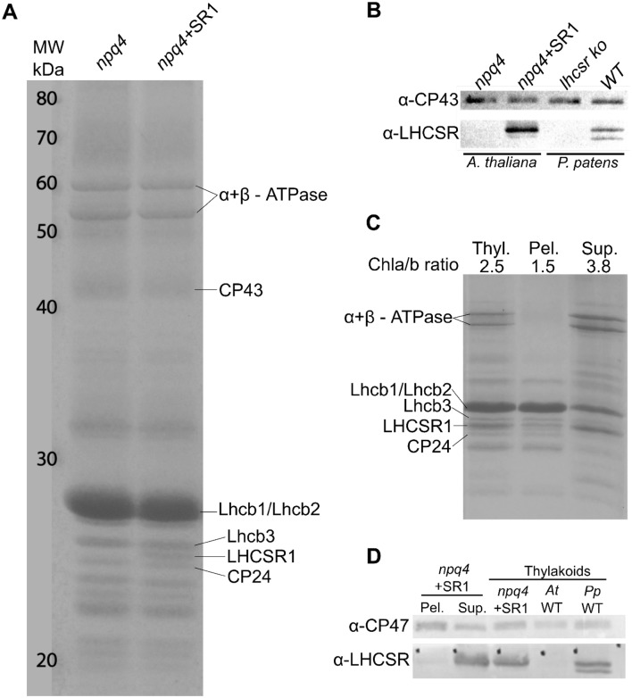
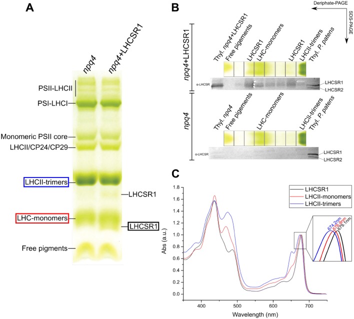
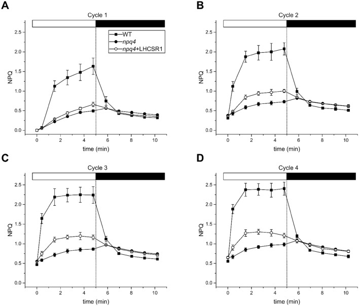
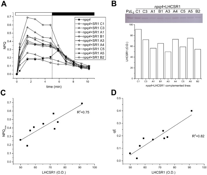
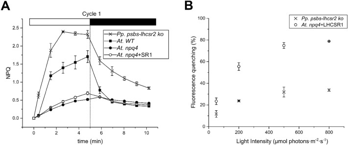
Non-photochemical quenching, NPQ, of chlorophyll fluorescence regulates the heat dissipation of chlorophyll excited states and determines the efficiency of the oxygenic photosynthetic systems. NPQ is regulated by a pH-sensing protein, responding to the chloroplast lumen acidification induced by excess light, coupled to an actuator, a chlorophyll/xanthophyll subunit where quenching reactions are catalyzed. In plants, the sensor is PSBS, while the two pigment-binding proteins Lhcb4 (also known as CP29) and LHCII are the actuators. In algae and mosses, stress-related light-harvesting proteins (LHCSR) comprise both functions of sensor and actuator within a single subunit. Here, we report on expressing the lhcsr1 gene from the moss Physcomitrella patens into several Arabidopsis thaliana npq4 mutants lacking the pH sensing PSBS protein essential for NPQ activity. The heterologous protein LHCSR1 accumulates in thylakoids of A. thaliana and NPQ activity can be partially restored. Complementation of double mutants lacking, besides PSBS, specific xanthophylls, allowed analyzing chromophore requirement for LHCSR-dependent quenching activity. We show that the partial recovery of NPQ is mostly due to the lower levels of Zeaxanthin in A. thaliana in comparison to P. patens. Complemented npq2npq4 mutants, lacking besides PSBS, Zeaxanthin Epoxidase, showed an NPQ recovery of up to 70% in comparison to A. thaliana wild type. Furthermore, we show that Lutein is not essential for the folding nor for the quenching activity of LHCSR1. In short, we have developed a system to study the function of LHCSR proteins using heterologous expression in a variety of A. thaliana mutants.

Keywords: A. thaliana; Heterologous expression; LHCSR; NPQ; P. patens; Photoprotection.


Conflict of interest statement

The authors declare that they have no conflict of interest.

Figures

Fig. 1
Fig. 1
A. thaliana thylakoid membrane fractionation and analysis of LHCSR distribution in the individual fractions. a Coomassie-stained SDS-PAGE separation of thylakoid proteins isolated from npq4 plants and npq4 plants expressing LHCSR1 (npq4 + SR1). LHCSR1 and other bands are indicated on the right side of the gel. b Western blot analysis of thylakoid proteins isolated from A. thaliana npq4 plants and npq4 plants expressing LHCSR. As control, thylakoids from P. patens WT and lhcsr KO were loaded. c Coomassie-stained SDS-PAGE of fractionated thylakoid membranes from A. thaliana npq4 + LHCSR1 using 0.47% α-DM. Thylakoid membranes (Thyl.), pellet enriched in grana fractions (Pel.) and the supernatant enriched in stroma membranes (Sup.). Gels were loaded on Chl basis, 4 µg for thylakoids and 2.7 µg for both the pellet and supernatant fractions. The Chl a/b ratio is indicated above the gel. d western blot analysis of the fractionated thylakoid membranes and thylakoids from A. thaliana npq4, npq4 + LHCSR1, WT and P. patens WT as controls
Fig. 2
Fig. 2
Deriphat-PAGE analysis of A. thaliana thylakoid membrane protein complexes. a Thylakoid membranes (30 μg of Chl) of npq4 and npq4 + LHCSR1 plants solubilized with 0.8% (w/v) α-DM were subjected to Deriphat-PAGE. PSII and PSI complexes, together with various LHCs are indicated on the left side of the gel. Complexes more abundant in npq4 + LHCSR1 than in npq4 plants are labeled as LHCSR1 on the right side of the gel. Colored rectangles correspond to the bands used for the absorption spectra analysis in (panel c). b Deriphat-PAGE (7%) of unstacked thylakoids from A. thaliana WT and npq4 + LHCSR1, solubilized in 0.8% α-DM. Bands were eluted in 10 mM HEPES/0.03% α-DM. Eluted fractions were loaded on SDS-PAGE and immunoblotted against α-LHCSR. c Absorption spectra of the bands taken from the Deriphat-PAGE (panel a), LHCSR1, LHCII-monomers and LHCII-trimers of A. thaliana npq4 + LHCSR1
Fig. 3
Fig. 3
NPQ kinetics (n = 4) in A. thaliana WT, npq4 and npq4 + LHCSR1. WT (Black squares), npq4 (black circles) and npq4 + LHCSR1 (open circles). Plants were dark adapted for 45 min before the measurement, 4 cycles of 5 min actinic light (1200 µmol photons m−2 s−1) and 5 min dark, as described in the M&M. The four cycles are depicted by ad, respectively
Fig. 4
Fig. 4
Correlation between NPQ activity and LHCSR1 accumulation. NPQ measurements of the T2 generation of the npq4 + LHCSR1 lines (n = 9). a. Leaves were dark adapted for 45 min, pre-treated with 1200 µmol photons m−2 s−1 of actinic light for 15 min and left to relax in the dark for 10 min before the NPQ measurement b After the NPQ measurement total leaf extracts from each line were loaded on an SDS-PAGE on a basis of 0.75 µg Chl and immunο-blotted against α-LHCSR antibodies. Thylakoids from P. patens psbs-lhcsr2 ko (PzL2) were loaded as a control. The O.D. of LHCSR1 was determined. c Protein level plotted with the maximum NPQ, yielding a positive correlation of R2 = 0.75. d Correlation between qE and the protein level (R2 = 0.82). qE recovery is calculated as the NPQ of the last point in the light phase minus the second point in the dark phase
Fig. 5
Fig. 5
Comparison of ΔpH and NPQ between P. patens psbs-lhcsr2 ko and A. thaliana.a Comparison of NPQ (n = 4) between A. thaliana WT, npq4, npq4 + LHCSR1 (npq4 + SR1) and P. patens psbs-lhcsr2 ko. Fourth cycle of NPQ measurements at 1200 µmol photons m−2 s−1 (5 min light and 5 min dark relaxation). b 9-aminoacridin measurements (n = 3) in isolated chloroplasts of A. thaliana npq4 + LHCSR1 and P. patens psbs-lhcsr2 ko at different light intensities (50, 200, 500 and 800 µmol photons m−2 s−1 of red light)
Fig. 6
Fig. 6
NPQ activity of npq4 + LHCSR1 lines in various light intensities. Three different A. thaliana npq4 + LHCSR1 lines with high and intermediate NPQ activation (line C1, A1 and A5) were tested in a variety of actinic light intensities. Leaves (n = 3) were dark adapted for 45 min, pre-treated with 800 µmol photons m−2 s−1 of actinic light for 15 min and left to relax in the dark for 10 min before the NPQ measurement. Each measurement corresponds to one single NPQ cycle of 5 min different with different actinic light intensities and 5 min dark recovery. The actinic light intensities used were: 100, 200, 400, 600, 800 and 1000 µmol photons m−2 s−1 (µE) from af, respectively. Leaves from A. thaliana WT and npq4 were used as controls. g NPQ of the last point in the light plotted against the different light intensities
Fig. 7
Fig. 7
NPQ measurements in A. thaliana mutants, lacking specific xanthophyll’s, complemented with LHCSR1. Four successive NPQ cycles were measured (n = 3) as described in M&M. The first and fourth cycle are shown for each complemented mutant. a, b first and fourth NPQ measurement in npq1np4 and three independent npq1npq4 lines complemented with LHCSR1 (npq1npq4 + SR1). c, d first and fourth NPQ measurement in npq2npq4 and three npq2npq4 lines complemented with LHCSR1 (npq2npq4 + SR1). e, f first and fourth NPQ measurement in lut2npq4 and three lut2npq4 lines complemented with LHCSR1 (lut2npq4 + SR1)

Similar articles

Cited by

References

    1. Ahn TK, Avenson TJ, Ballottari M, et al. Architecture of a charge-transfer state regulating light harvesting in a plant antenna protein. Science. 2008;320:794–797. doi: 10.1126/science.1154800. - DOI - PubMed
    1. Alboresi A, Gerotto C, Giacometti GM, et al. Physcomitrella patens mutants affected on heat dissipation clarify the evolution of photoprotection mechanisms upon land colonization. Proc Natl Acad Sci USA. 2010;107:11128–11133. doi: 10.1073/pnas.1002873107. - DOI - PMC - PubMed
    1. Allorent G, Tokutsu R, Roach T, et al. A dual strategy to cope with high light in Chlamydomonas reinhardtii. Plant Cell. 2013;25:545–557. doi: 10.1105/tpc.112.108274. - DOI - PMC - PubMed
    1. Ashton NW, Grimsley NH, Cove DJ. Analysis of gametophytic development in the moss, Physcomitrella patens, using auxin and cytokinin resistant mutants. Planta. 1979;144:427–435. doi: 10.1007/BF00380118. - DOI - PubMed
    1. Baker NR. Chlorophyll fluorescence: a probe of photosynthesis in vivo. Annu Rev Plant Biol. 2008;59:89–113. doi: 10.1146/annurev.arplant.59.032607.092759. - DOI - PubMed

MeSH terms

LinkOut - more resources