

Inhibition of Reactive Astrocytes with Fluorocitrate Ameliorates Learning and Memory Impairment Through Upregulating CRTC1 and Synaptophysin in Ischemic Stroke Rats
- PMID: 31270712
- PMCID: PMC11452224
- DOI: 10.1007/s10571-019-00709-0
Inhibition of Reactive Astrocytes with Fluorocitrate Ameliorates Learning and Memory Impairment Through Upregulating CRTC1 and Synaptophysin in Ischemic Stroke Rats
Abstract
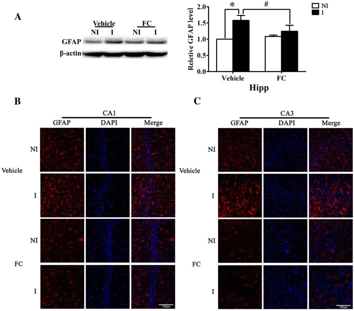
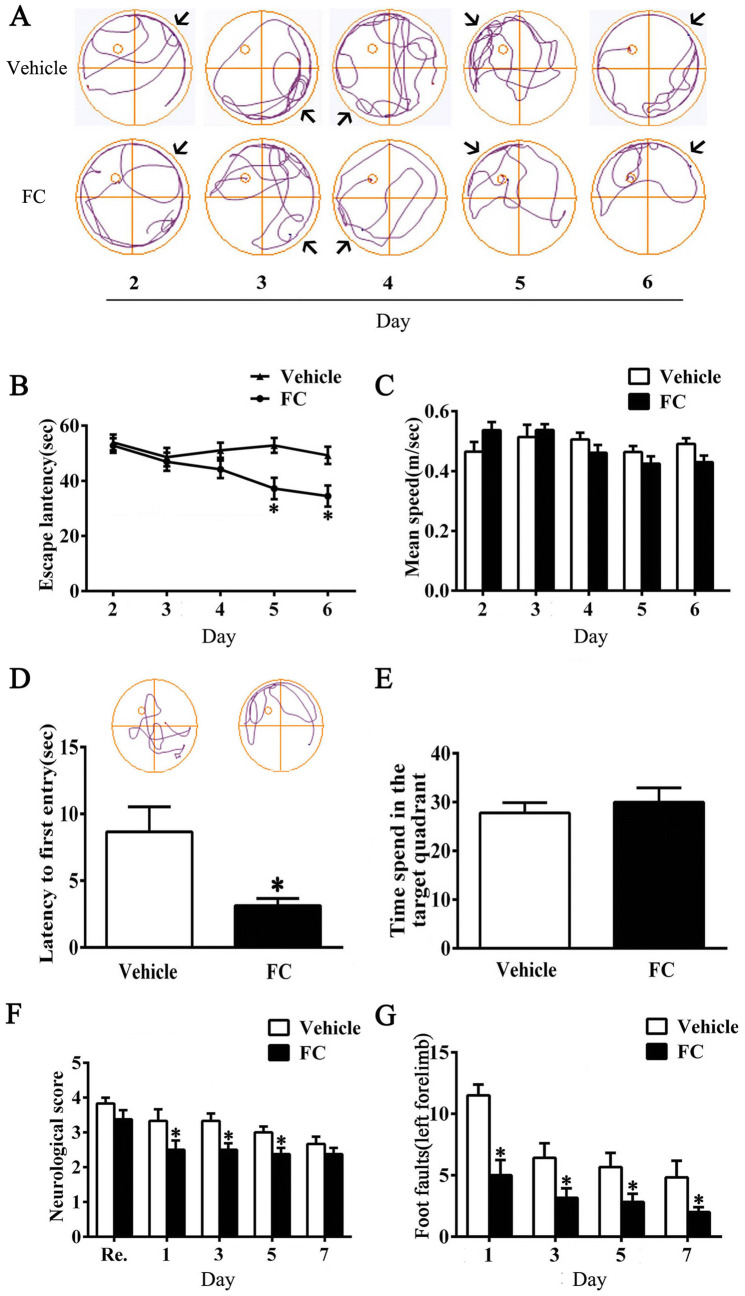
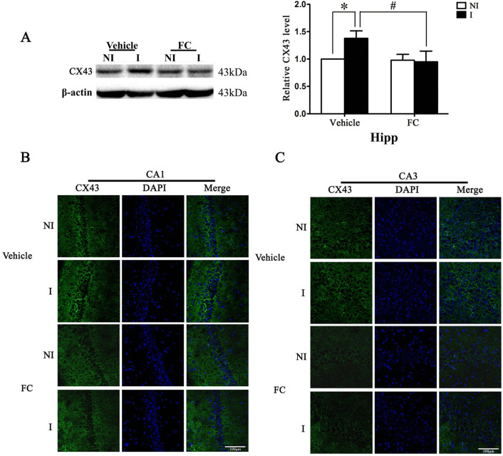
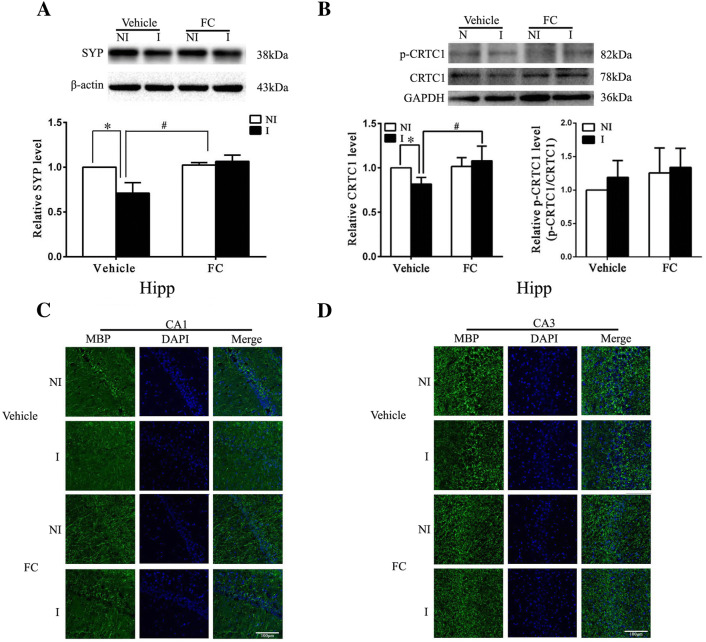
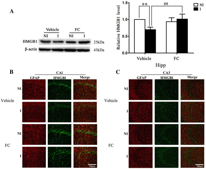
Ischemic stroke often causes motor and cognitive deficits. Deregulated glia gap junction communication, which is reflected by increased protein levels of glial fibrillary acidic protein (GFAP) and connexin 43 (Cx43), has been observed in ischemic hippocampus and has been associated with cognitive impairment in animal stroke models. Here, we tested the hypothesis that reactive astrocytes-mediated loss of synaptophysin (SYP) and CREB-regulated transcription coactivator 1 (CRTC1) contribute to dysfunction in glia gap junction communication and memory impairment after ischemic stroke. Male Sprague-Dawley rats were subjected to a 90-min middle cerebral artery occlusion (MCAO) with 7-day reperfusion. Fluorocitrate (1 nmol), the reversible inhibitor of the astrocytic tricarboxylic acid cycle, was injected into the right lateral ventricle of MCAO rats once every 2 days starting immediately before reperfusion. The Morris water maze was used to assess memory in conjunction with western blotting and immunostaining to detect protein expression and distribution in the hippocampus. Our results showed that ischemic stroke caused significant memory impairment accompanied by increased protein levels of GFAP and Cx43 in hippocampal tissue. In addition, the levels of several key memory-related important proteins including SYP, CRTC1, myelin basic protein and high-mobility group-box-1 were significantly reduced in the hippocampal tissue. Of note, inhibition of reactive astrocytes with fluorocitrate was shown to significantly reverse the above noted changes induced by ischemic stroke. Taken together, our findings demonstrate that inhibiting reactive astrocytes with fluorocitrate immediately before reperfusion may protect against ischemic stroke-induced memory impairment through the upregulation of CRTC1 and SYP.
Keywords: CRTC1; Cx43; Fluorocitrate; Ischemic stroke; Memory; Reactive astrocytes.
Conflict of interest statement
The authors declare that they have no conflict of interest.
Figures

Similar articles
-
Resveratrol Attenuates Subacute Systemic Inflammation-Induced Spatial Memory Impairment via Inhibition of Astrocyte Activation and Enhancement of Synaptophysin Expression in the Hippocampus.Ann Clin Lab Sci. 2017 Jan;47(1):17-24. Ann Clin Lab Sci. 2017. PMID: 28249911
-
Bone marrow stromal cells upregulate expression of bone morphogenetic proteins 2 and 4, gap junction protein connexin-43 and synaptophysin after stroke in rats.Neuroscience. 2006 Aug 25;141(2):687-695. doi: 10.1016/j.neuroscience.2006.04.054. Epub 2006 May 30. Neuroscience. 2006. PMID: 16730912
-
Inhibition of reactive astrocytes with fluorocitrate retards neurovascular remodeling and recovery after focal cerebral ischemia in mice.J Cereb Blood Flow Metab. 2010 Apr;30(4):871-82. doi: 10.1038/jcbfm.2009.257. Epub 2009 Dec 9. J Cereb Blood Flow Metab. 2010. PMID: 19997116 Free PMC article.
-
A novel cognitive impairment mechanism that astrocytic p-connexin 43 promotes neuronic autophagy via activation of P2X7R and down-regulation of GLT-1 expression in the hippocampus following traumatic brain injury in rats.Behav Brain Res. 2015 Sep 15;291:315-324. doi: 10.1016/j.bbr.2015.05.049. Epub 2015 May 29. Behav Brain Res. 2015. PMID: 26031379
-
Remote ischemic post-conditioning improves neurological function by AQP4 down-regulation in astrocytes.Behav Brain Res. 2015 Aug 1;289:1-8. doi: 10.1016/j.bbr.2015.04.024. Epub 2015 Apr 21. Behav Brain Res. 2015. PMID: 25907740 Review.
Cited by
-
New Insights Into the Pivotal Role of CREB-Regulated Transcription Coactivator 1 in Depression and Comorbid Obesity.Front Mol Neurosci. 2022 Feb 15;15:810641. doi: 10.3389/fnmol.2022.810641. eCollection 2022. Front Mol Neurosci. 2022. PMID: 35242012 Free PMC article. Review.
-
A novel rhein-huprine hybrid ameliorates disease-modifying properties in preclinical mice model of Alzheimer's disease exacerbated with high fat diet.Cell Biosci. 2023 Mar 9;13(1):52. doi: 10.1186/s13578-023-01000-y. Cell Biosci. 2023. PMID: 36895036 Free PMC article.
-
In Vitro Astroglial Dysfunction Induced by Neurotoxins: Mimicking Astrocytic Metabolic Alterations of Alzheimer's Disease.Metabolites. 2024 Mar 1;14(3):151. doi: 10.3390/metabo14030151. Metabolites. 2024. PMID: 38535311 Free PMC article.
-
Silencing of Long Noncoding RNA GAS5 Blocks Experimental Cerebral Ischemia-Reperfusion Injury by Restraining AQP4 Expression via the miR-1192/STAT5A Axis.Mol Neurobiol. 2022 Dec;59(12):7450-7465. doi: 10.1007/s12035-022-03045-5. Epub 2022 Oct 5. Mol Neurobiol. 2022. PMID: 36195691
-
Peroxisomal Proliferator-Activated Receptor β/δ Deficiency Induces Cognitive Alterations.Front Pharmacol. 2022 Jul 11;13:902047. doi: 10.3389/fphar.2022.902047. eCollection 2022. Front Pharmacol. 2022. PMID: 35899125 Free PMC article.
References
-
- Adamsky A, Goshen I (2018) Astrocytes in memory function: pioneering findings and future directions. Neuroscience 370:14–26 - PubMed
-
- Bakker FC, Klijn CJ, Jennekens-Schinkel A, Kappelle LJ (2000) Cognitive disorders in patients with occlusive disease of the carotid artery: a systematic review of the literature. J Neurol 247(9):669–676 - PubMed
-
- Baron JC (2018) Protecting the ischaemic penumbra as an adjunct to thrombectomy for acute stroke. Nat Rev Neurol 14(6):325–337 - PubMed
MeSH terms
Substances
Grants and funding
LinkOut - more resources
Full Text Sources
Medical
Miscellaneous
