. 2019;56(5):217-229.
doi: 10.1159/000501312. Epub 2019 Jul 4.

Effects of Iliac Stenosis on Abdominal Aortic Aneurysm Formation in Mice and Humans

Affiliations

Effects of Iliac Stenosis on Abdominal Aortic Aneurysm Formation in Mice and Humans

Gurneet S Sangha et al. J Vasc Res. 2019.

Abstract

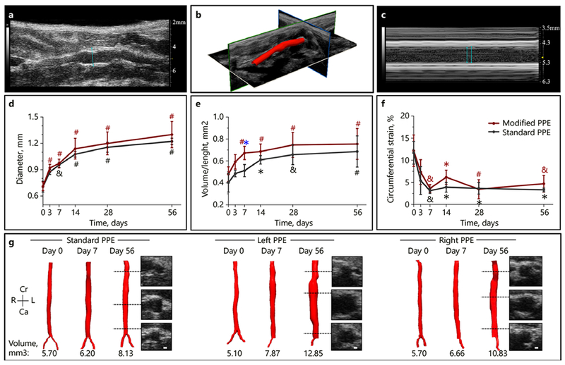
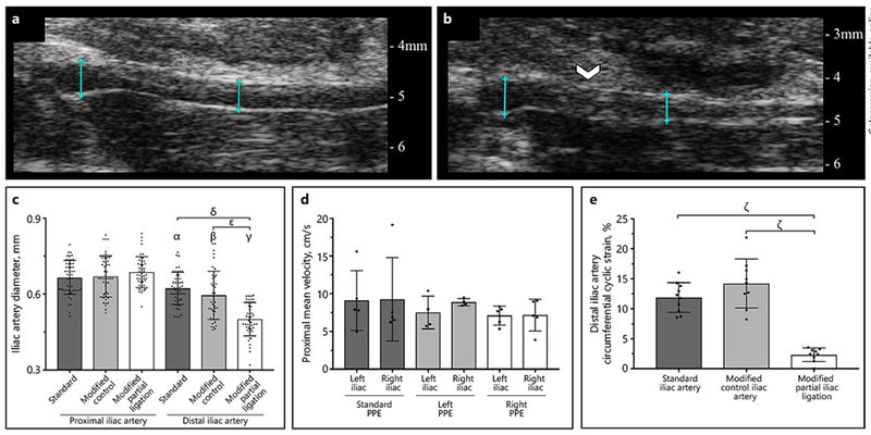
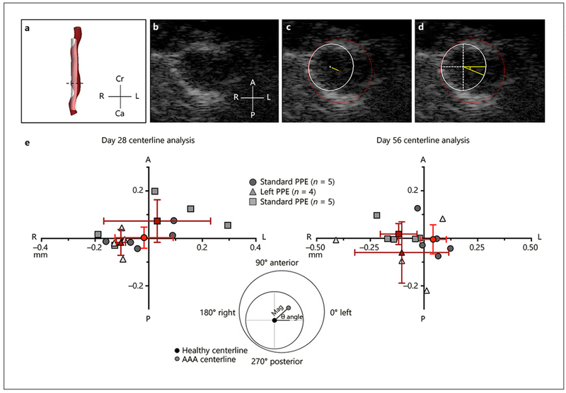
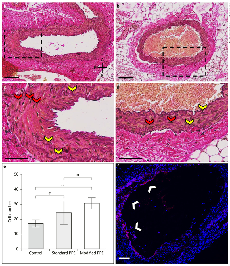
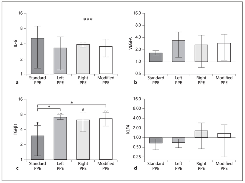
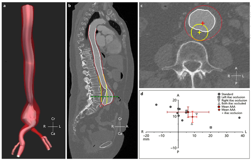
Reduced lower-limb blood flow has been shown to lead to asymmetrical abdominal aortic aneurysms (AAAs) but the mechanism of action is not fully understood. Therefore, small animal ultrasound (Vevo2100, FUJIFILM VisualSonics) was used to longitudinally study mice that underwent standard porcine pancreatic elastase (PPE) infusion (n = 5), and PPE infusion with modified 20% iliac artery stenosis in the left (n = 4) and right (n = 5) iliac arteries. Human AAA computed tomography images were obtained from patients with normal (n = 9) or stenosed left (n = 2), right (n = 1), and bilateral (n = 1) iliac arteries. We observed rapid early growth and rightward expansion (8/9 mice) in the modified PPE groups (p < 0.05), leading to slightly larger and asymmetric AAAs compared to the standard PPE group. Further examination showed a significant increase in TGFβ1 (p < 0.05) and cellular infiltration (p < 0.05) in the modified PPE group versus standard PPE mice. Congruent, yet variable, observations were made in human AAA patients with reduced iliac outflow compared to those with normal iliac outflow. Our results suggest that arterial stenosis at the time of aneurysm induction leads to faster AAA growth with aneurysm asymmetry and increased vascular inflammation after 8 weeks, indicating that moderate iliac stenosis may have upstream effects on AAA progression.

Keywords: Aneurysm; Asymmetry; Iliac artery; Murine.


Conflict of interest statement

Disclosure Statement

The authors have no conflict of interest to declare.

Figures

Fig. 1.
Fig. 1.
Representative healthy (left panels) and aneurysmal (right panels) murine aortae with B-mode (a, b), M-mode (c, d), and PWD (e, f) images. B-mode images were used to assess overall vessel structure and quantify the increase in aortic diameter after PPE treatment. M-mode was used to measure circumferential cyclic strain, and PWD was used to quantify mean and peak velocities.
Fig. 2.
Fig. 2.
Summary of structural changes in murine abdominal aortic aneurysm development among standard, left, and right PPE groups. Long-axis B-mode was used to quantify aortic diameter (a), 3D ultrasound for aortic segmentation and volume characterization (b), and M-mode to quantify systolic and diastolic diameters (c) that were used to assess changes in circumferential cyclic strain. The aortic diameter continues to increase up to day 56 (d) with the modified PPE group having a rapid increase in volume/length up to day 7 (e). We also observed a sharp decrease in circumferential cyclic strain between baseline and day 7 post-procedure time points (f). Representative short-axis ultrasound, 3D segmentation, and volume showed slightly larger aneurysm in the modified PPE group compared to the standard PPE group (g). Statistical significance compared with day 0 and defined at * p < 0.05, & p < 0.01, and # p < 0.001. The blue asterisk (e) represents statistical significance between the standard and modified PPE groups.
Fig. 3.
Fig. 3.
Comparison of structural and hemodynamic changes between non-ligated (a) and partially ligated (b) iliac arteries at day 56. We observed a significant decrease in diameter (c) and circumferential cyclic strain (e) in modified iliac arteries compared to standard PPE arteries and contralateral control iliac arteries. We did not, however, see significant changes in iliac artery velocity between standard and modified PPE groups at day 56 (d). Suture placement is highlighted by the white arrow and diameter measurement locations are shown by blue lines. Statistical significance is shown as p < 0.001 (α, β, γ, δ, ε, ζ). Symbols α, β, γ signify statistical significance between their respective group and the standard, modified control, and modified partial ligation groups in the proximal iliac artery.
Fig. 4.
Fig. 4.
Summary of murine centerline deviation analysis methods and results. Aneurysm and projected healthy regions were first segmented using SimVascular (a). We then used short-axis view (b) to quantify magnitude (c) and direction (d) of centerline deviation between the healthy and diseased aorta. Centerline deviation results (e) for day 28 and 56 shows the shift in magnitude and direction of the diseased vessels with respect to the healthy vessel. Red points represent the mean centerline deviation for the standard and modified PPE groups, and the red lines represent the standard deviation in the x and y axes. Cr, cranial; Ca, caudal; L, left; R, right; A, anterior; P, posterior.
Fig. 5.
Fig. 5.
Histological and immunohistochemistry analysis of PPE-infused aortic tissue consisting of EvG-stained standard (a, c) and modified (b, d) aortae, as well as Ki67-stained modified (f) infrarenal aorta. Histology shows diffuse elastin breakage throughout the vessels (yellow arrows), thus confirming aneurysm induction. Ki67 staining shows cell proliferation in the standard and modified PPE animals (white arrows). Further, cell number analysis showed a significant increase in cell nuclei (red arrows) between the modified and standard PPE-infused aortae (h). A, anterior; P, posterior; L, left; R, right. Scale bars = 100 μm (a, c) and 50 μm (b, d). Statistical significance is defined at * p < 0.05, # p < 0.001, ~ p < 0.0001.
Fig. 6.
Fig. 6.
Relative gene expression analysis of standard and modified infrarenal aortae at day 56 using qPCR. IL-6 (a) and VEGFA (b) are upregulated, but there are no significant differences between PPE-infused and control aortae. TGFβ1 shows significant upregulation between PPE-infused and control aortae, and standard and modified groups (c); while KLF4 (d) shows variable differences in expression between standard and modified groups. Fold change values are relative to control aortae. Statistical significance is defined at * p < 0.05, # p < 0.001, and ~ p < 0.0001.
Fig. 7.
Fig. 7.
Summary of human centerline deviation analysis methods and results. 3D segmentation was performed on human AAA (a) and a sagittal cross-sectional plane was used to confirm quality of 3D segmentation with red, white, and yellow highlighting the AAA wall, AAA lumen, and projected healthy aorta, respectively (b). A transverse cross-sectional plane along the center of the AAA (green dashed line) was used to quantify magnitude and direction of centerline deviation (c). Quantitative analysis shows that standard and modified AAA grew away from the spine in the leftward direction, likely due to the natural leftward origin of the healthy aorta. Red points represent the mean centerline deviation for the standard and modified PPE groups, and the red lines represent the standard deviation in the x and y axes. Cr, cranial; Ca, caudal; L, left; R, right; A, anterior; P, posterior.

References

    1. Sampson UK, Norman PE, Fowkes FG, Aboyans V, Song Y, Harrell FE Jr, et al. Estimation of global and regional incidence and prevalence of abdominal aortic aneurysms 1990 to 2010. Glob Heart. 2014. March;9(1):159–70. - PubMed
    1. Sampson UKA, Norman PE, Fowkes FGR, Aboyans V, Song Y, Harrell FE Jr, et al. Global and regional burden of aortic dissection and aneurysms: mortality trends in 21 world regions, 1990 to 2010. Glob Heart. 2014 Mar; 9(1):171–80.e10. - PubMed
    1. Vardulaki KA, Prevost TC, Walker NM, Day NE, Wilmink AB, Quick CR, et al. Incidence among men of asymptomatic abdominal aortic aneurysms: estimates from 500 screen detected cases. J Med Screen. 1999;6(1):50–4. - PubMed
    1. Wassef M, Baxter BT, Chisholm RL, Dalman RL, Fillinger MF, Heinecke J, et al. Pathogenesis of abdominal aortic aneurysms: a multidisciplinary research program supported by the National Heart, Lung, and Blood Institute. J Vasc Surg. 2001. October;34(4):730–8. - PubMed
    1. Vollmar JF, Paes E, Pauschinger P, Henze E, Friesch A. Aortic aneurysms as late sequelae of above-knee amputation. Lancet. 1989. October; 2(8667):834–5. - PubMed

Publication types

MeSH terms

Substances