. 2019 Aug 21;103(4):583-597.e8.
doi: 10.1016/j.neuron.2019.05.047. Epub 2019 Jul 1.

Plug-and-Play Protein Modification Using Homology-Independent Universal Genome Engineering

Affiliations

Plug-and-Play Protein Modification Using Homology-Independent Universal Genome Engineering

Yudong Gao et al. Neuron. .

Abstract

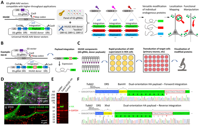
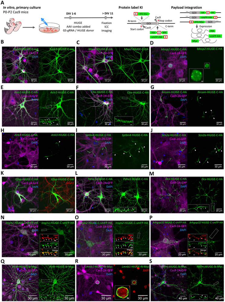
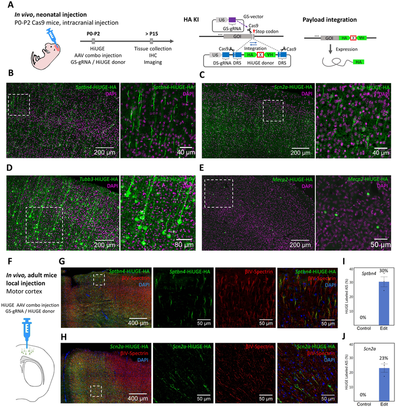
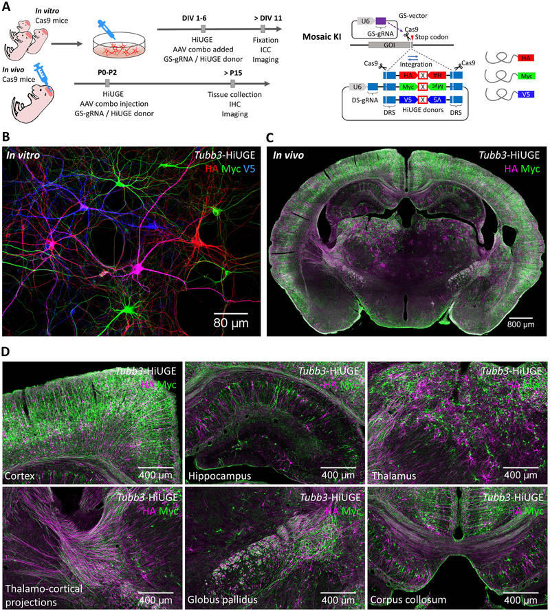
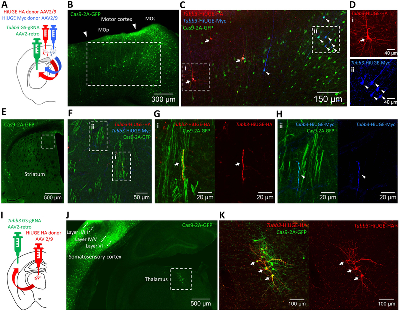
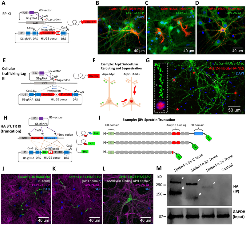
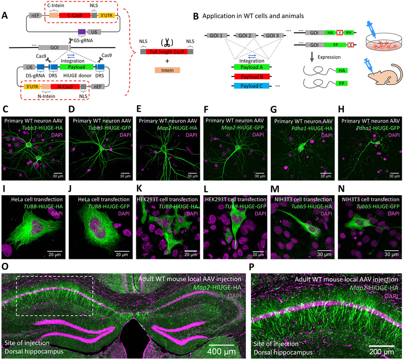
Analysis of endogenous protein localization, function, and dynamics is fundamental to the study of all cells, including the diversity of cell types in the brain. However, current approaches are often low throughput and resource intensive. Here, we describe a CRISPR-Cas9-based homology-independent universal genome engineering (HiUGE) method for endogenous protein manipulation that is straightforward, scalable, and highly flexible in terms of genomic target and application. HiUGE employs adeno-associated virus (AAV) vectors of autonomous insertional sequences (payloads) encoding diverse functional modifications that can integrate into virtually any genomic target loci specified by easily assembled gene-specific guide-RNA (GS-gRNA) vectors. We demonstrate that universal HiUGE donors enable rapid alterations of proteins in vitro or in vivo for protein labeling and dynamic visualization, neural-circuit-specific protein modification, subcellular rerouting and sequestration, and truncation-based structure-function analysis. Thus, the "plug-and-play" nature of HiUGE enables high-throughput and modular analysis of mechanisms driving protein functions in cellular neurobiology.

Keywords: CRISPR; HiUGE; genomics; immunolabeling; knockin; proteomics.


Conflict of interest statement

Declaration of Interests

S.H.S. and Y.G. have filed a patent application related to this work.

S.H.S. is a founder of CasTag Biosciences and a member of its scientific advisory board.

Figures

Figure 1.
Figure 1.. Illustration of the HiUGE system.
(A) Schematic of HiUGE method. The HiUGE donor vector expresses a donor-specific gRNA (DS-gRNA), which specifically recognizes the donor recognition sequence (DRS) and directs the cleavage and release of the donor payload for insertion into gene-specific gRNA (GS-gRNA) targeted loci for modification of proteins. (B) HiUGE donor vectors harboring short epitope tags employ a dual-orientation design for efficient expression following either forward or reverse genomic integration. A cassette of stop codons for all six ORFs (denoted by an “X” boxed in red, sequence listed in Table S1) is used to terminate translation. (C) Workflow using a small-scale, low-cost, and higher-throughput AAV supernatant production method for in vitro applications. (D-F) Proof-of-principle data showing HA-epitope KI to mouse Tubb3 gene in primary neuron culture of Cas9 mice after transduction with a combination of small-scale GS-gRNA AAV and HA-epitope donor AAV. (D) Representative (i) confocal and (ii) stimulated emission depletion (STED) images of immunocytochemistry staining for HA-epitope, showing the characteristic microtubule expression pattern of the HA-epitope labeled ßIII-tubulin. GFP fluorescence of the Cas9-2A-GFP, nuclei labeling with DAPI (4',6-diamidino-2-phenylindole), and synaptic marker Synapsin I staining are also shown. (E) Western blot for HA-epitope in a comparison against a negative control (transduced with empty GS-gRNA backbone and HiUGE donor), showing a single band of labeled protein at the expected molecular mass (~51kD) for ßIII-tubulin. (F) Representative sequencing results showing correct integration in forward and reverse orientation. Regions of DNA sequence containing genomic Tubb3, DRS (blue shade), restriction enzymes (yellow shade), HA-epitope (green shade) and stop codon cassette (red box) are indicated. Truncated terminal amino acids are noted. Translated amino acid sequences encoded by the payload and the in-frame stop codon (red asterisk) are indicated as well.
Figure 2.
Figure 2.. Rapid Protein Modification Across Diverse Genomic and Protein Targets with HiUGE In Vitro.
(A) Schematic of HiUGE KI application for C- or N-term protein labeling in vitro. Primary hippocampal cells from Cas9 mice were transduced with a combination of GS-gRNA and HiUGE donor AAVs and immunostained to detect epitope or smFP-HA labeling, with representative images displayed in panels B-S. (B-M) Examples of C-term HA-epitope KI to diverse targets (mouse Tubb3, Map2, Mecp2, Actr2, Clta, Nrcam, Ank3, Sptbn4, Scn2a, Gfap, Pdha1, and Dcx), showing the expected expression patterns of the translated proteins respectively. (N-P) C-term smFP-HA KI to mouse Insyn1, Insyn2, and Arhgap32, which encode the inhibitory postsynaptic density (iPSD) proteomic candidates. Colocalization of the HA-immunoreactivity with the juxtaposed inhibitory presynaptic marker vesicular GABA transporter (VGAT) immunosignal is shown in the insets. (Q-S) N-term Myc-epitope KI to mouse Actb, Lmnb1, and Nefm, showing the expected expression patterns of the translated proteins respectively. Scale bar is indicated in each panel, or within insets (2μm). GFP fluorescence of the Cas9-2A-GFP and nuclei labeling with DAPI are also shown. Arrowheads represent the subcellular features associated with the targeted genes, such as the dendritic spines, AIS, mitochondria, distal end of neurites, inhibitory synapses, and neurofilaments.
Figure 3.
Figure 3.. Endogenous Protein Labeling with HiUGE In Vivo.
(A) Schematic of C-term HA-epitope KI in vivo. Neonatal Cas9 pups were intracranially injected with a combination of purified GS-gRNA AAV and HiUGE donor AAV, and euthanized after P15 for immunohistochemistry to detect HA-epitope KI. (B-E) Representative images of HA-epitope KI to mouse Sptbn4, Scn2a, Tubb3, and Mecp2 gene, showing the expected expression patterns of the translated proteins respectively. Cortical regions of coronal sections are shown. Image on the right within each panel shows a magnified view of the boxed region from the left image. (F) Adult Cas9 mice were locally injected with a combination of purified GS-gRNA and HA-epitope donor AAVs (titer of 3 × 10¹⁰ GC / μL) into the motor cortex and euthanized 2 weeks later for immunohistochemistry. (G-H) Representative immunofluorescent images of HA-epitope (green) following HiUGE-mediated KI to mouse Sptbn4 and Scn2a. Antibody labeling of the AIS-marker (ßIV-spectrin) is also shown (red). (I-J) Quantification of cellular labeling efficiency showing the fractions of the ßIV-spectrin-positive AIS-structures labeled with HiUGE at the injection sites (Sptbn4: 30.2 ± 3.4 %; Scn2a: 22.8 ± 2.9 %, n=3 mice), in comparison to negative controls (uninjected, 0%). Error bars represent standard error of the mean (SEM). Scale bar is indicated in each panel.
Figure 4.
Figure 4.. HiUGE Donor Payload Interchangeability for Multiplexed Protein Modification.
(A) Schematic of C-term KI of epitope mixture in vitro and in vivo. (B) Labeling of HA, Myc, and V5-epitope following mosaic KI to mouse Tubb3, showing stochastically integrated epitopes in neighboring neurons in vitro. Occasionally, cells positive for two epitopes can be seen (e.g. the magenta-colored cell in this image, showing both HA and V5 immunoreactivity). (C) Coronal section demonstrating labeling of HA and Myc-epitope following mosaic KI to mouse Tubb3 in vivo. (D) Zoomed images showing the cortex, hippocampus, thalamus, thalamo-cortical projections, globus pallidus, and corpus collosum of panel (C). Scale bar is indicated in each panel.
Figure 5.
Figure 5.. Neural Circuit-based HiUGE Labeling.
(A) Illustration of cortico-striatal circuit-selective C-term labeling of ßIII-tubulin by injection of AAV2-retro mouse Tubb3 GS-gRNA into the striatum and 2 lateral injections of AAV2/9 HA and Myc-epitope donors in the motor cortex. (B) Representative image showing GFP labeling in the motor cortex, indicating retrogradely accessed Cre-dependent Cas9– 2A-GFP expression in projection neurons. (C) Immunolabeling of HA (arrows) and Myc-epitope (arrowheads) tagged ßIII-tubulin, imaged from the boxed area in (B). (D) Enlarged images from the boxed areas in (C), showing cells positive for (i) HA or (ii) Myc-epitope. (E) GFP signal from the AAV2-retro injected striatum. (F) Zoomed image of the boxed area in (E), showing GFP-positive axon bundles that contain fibers positive for HA or Myc-epitope. (G, H) Enlarged images showing fibers positive for (i) HA or (ii) Myc-epitope within GFP-positive axon bundles from boxed areas in (F). (I) Illustration of thalamo-cortical circuit-selective C-term labeling of ßIII-tubulin by injection of AAV2- retro mouse Tubb3 GS-gRNA in the somatosensory cortex and injection of AAV2/9 HA-epitope donor in the thalamus. (J) Representative image showing retrogradely activated Cas9-2A-GFP expression within the thalamus (boxed area) and local cortical networks (mostly cells within layer II/III and layer VI). (K) Zoomed image of the boxed area in (J), showing retrogradely accessed and HiUGE edited thalamic neurons positive for HA-epitope (arrows) and Cas9-2A-GFP. Scale bar is indicated in each panel.
Figure 6.
Figure 6.. HiUGE Payloads Enable Flexible Selection Across Multiple Methods to Interrogate Endogenous Protein Functions.
(A) Schematic of fluorescent protein (FP) KI with flexible (GGGGS)4 linker. (B-D) Detection of mCherry (mCh) KI to different genomic targets (mouse Tubb3, Gfap, and Pdha1) with (B) antibody against mCh, or (C, D) direct visualization of mCh fluorescence. (E) Schematic of HiUGE subcellular re-routing constructs using cellular trafficking tags as the payloads. (F) Example using a HiUGE payload with HA-epitope and nuclear localization signal (HA-NLS) to sequester an actin cytoskeletal protein Arp2 to the nucleus. (G) Representative image of immunostaining following C-term HA-NLS KI to mouse Actr2 gene, showing the HA-NLS-tagged Arp2 (red) redirected to the nucleus. Simultaneously, the Myc-epitope (no NLS) tagged Arp2 (green) was enriched at the dendritic spines, consistent with the normal localization of Arp2. (H) Schematic illustration of HiUGE HA-3’ untranslated region (3’-UTR) KI to truncate endogenous proteins for conducting structure-function relationship studies. (I) Example using HA-3’UTR KI strategy to truncate ßIV-spectrin, composed of calponin-homology (CH) domains, spectrin repeats, and a pleckstrin homology (PH) domain. (J) C-term KI using a GS-gRNA targeting near the stop codon of the last coding exon 36 (e.36) of ßIV-spectrin is compared to (K) truncation of PH domain by targeting exon 31 (e.31), which still retains AIS enrichment, or (L) disruption of spectrin repeat 14 and truncation of downstream sequences by targeting exon 26 (e.26), which completely disrupts the AIS localization. (M) Western blot of HA-epitope showing stepwise reduction of molecular mass consistent with the serial truncation conditions (arrowheads). Arrowheads indicate the Σ1 isoform of ßIV-spectrin, while arrows indicate the Σ6 isoform. The Σ6 isoform of the truncation at exon 26 (e.26) appeared undetectable. Molecular mass is marked in reference to the ladder. Scale bar is indicated in each panel.
Figure 7.
Figure 7.. HiUGE Vectors with Intein-mediated Split-Cas9 Trans-splicing.
(A) Schematic of the HiUGE system that harbors intein-mediated split-Cas9 expression for use in WT cells and animals. (B) Illustration of delivering interchangeable payloads to diverse genomic loci using these vectors. (C-H) Wild-type (WT) primary mouse neurons were AAV transduced and (I-N) common human or mouse cell lines were plasmid transfected with a combination of GS-gRNAs and HiUGE donors, followed by immunostaining for HA-epitope or GFP to detect payload KI. (C, E, G) Representative images of HA-epitope KI to the mouse Tubb3, Map2 and Pdha1 genes, showing the expected (C, E) microtubule and (G) mitochondrial localization of the tagged proteins. (D, F, H) Representative images of GFP KI to the mouse Tubb3, Map2 and Pdha1 genes, showing the expected (D, F) microtubule and (H) mitochondrial localization of the tagged proteins. (I, K, M) Representative images of HA-epitope KI to the human TUBB or mouse Tubb5 gene, showing the expected microtubule localization of the tagged proteins in (I) HeLa cell, (K) HEK293T cell and (M) NIH3T3 cell. (J, L, N) Representative images of GFP KI to the human TUBB or mouse Tubb5 gene, showing the expected microtubule localization of the tagged proteins in (J) HeLa cell, (L) HEK293T cell and (N) NIH3T3 cell. (O) Representative image of HA-epitope KI to mouse Map2 gene following local AAV injection in the dorsal hippocampus of adult WT mice, showing efficient labeling of the neurons at the injection site compared to negative labeling on the contralateral side. Zoomed view of the boxed area is shown in (P). Scale bar is indicated in each panel.

References

    1. Aurnhammer C, Haase M, Muether N, Hausl M, Rauschhuber C, Huber I, Nitschko H, Busch U, Sing A, Ehrhardt A, and Baiker A (2012). Universal real-time PCR for the detection and quantification of adeno-associated virus serotype 2-derived inverted terminal repeat sequences. Hum Gene Ther Methods 23, 18–28. - PubMed
    1. Bae S, Park J, and Kim JS (2014). Cas-OFFinder: a fast and versatile algorithm that searches for potential off-target sites of Cas9 RNA-guided endonucleases. Bioinformatics 30, 1473–1475. - PMC - PubMed
    1. Baker KE, and Parker R (2004). Nonsense-mediated mRNA decay: terminating erroneous gene expression. Curr Opin Cell Biol 16, 293–299. - PubMed
    1. Bear JE, Loureiro JJ, Libova I, Fassler R, Wehland J, and Gertler FB (2000). Negative regulation of fibroblast motility by Ena/VASP proteins. Cell 101, 717–728. - PubMed
    1. Berglund L, Bjorling E, Oksvold P, Fagerberg L, Asplund A, Szigyarto CA, Persson A, Ottosson J, Wernerus H, Nilsson P, et al. (2008). A genecentric Human Protein Atlas for expression profiles based on antibodies. Mol Cell Proteomics 7, 2019–2027. - PubMed

Publication types

MeSH terms

Substances