. 2019 Jul 5;365(6448):53-60.
doi: 10.1126/science.aau9263.

A COPII subunit acts with an autophagy receptor to target endoplasmic reticulum for degradation

Affiliations

A COPII subunit acts with an autophagy receptor to target endoplasmic reticulum for degradation

Yixian Cui et al. Science. .

Abstract

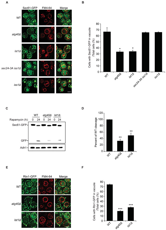
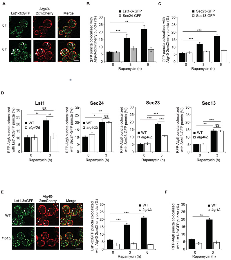
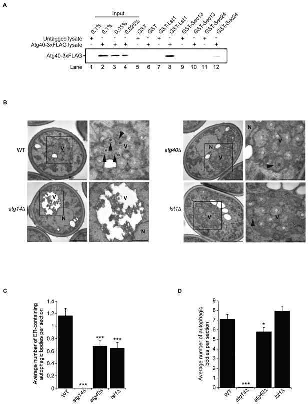
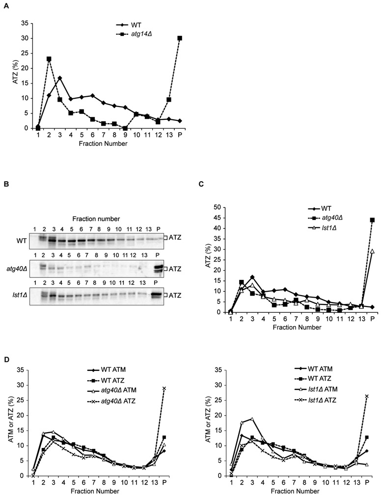
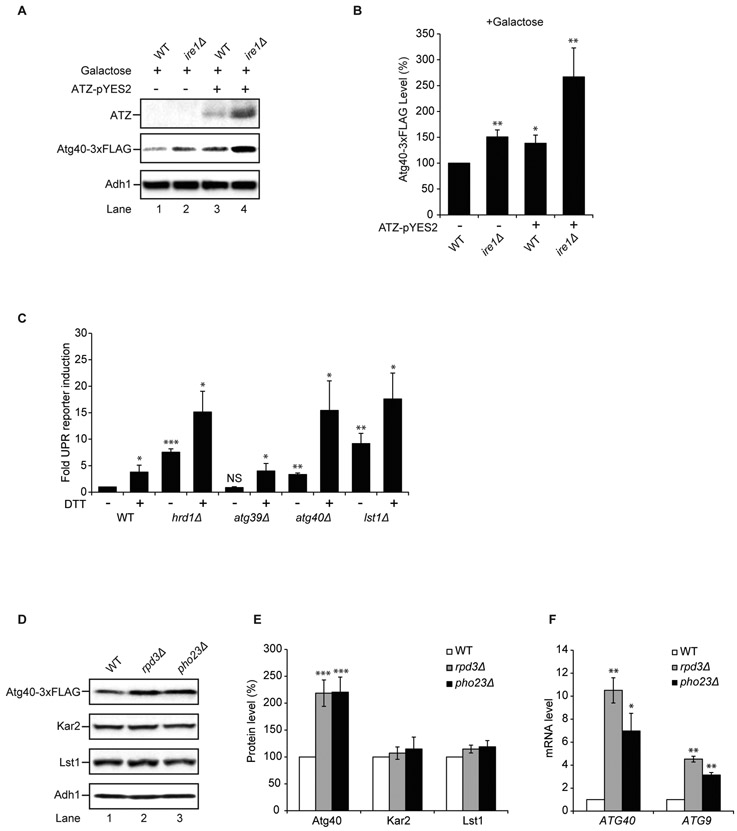
The COPII-cargo adaptor complex Lst1-Sec23 selectively sorts proteins into vesicles that bud from the endoplasmic reticulum (ER) and traffic to the Golgi. Improperly folded proteins are prevented from exiting the ER and are degraded. ER-phagy is an autophagic degradation pathway that uses ER-resident receptors. Working in yeast, we found an unexpected role for Lst1-Sec23 in ER-phagy that was independent from its function in secretion. Up-regulation of the stress-inducible ER-phagy receptor Atg40 induced the association of Lst1-Sec23 with Atg40 at distinct ER domains to package ER into autophagosomes. Lst1-mediated ER-phagy played a vital role in maintaining cellular homeostasis by preventing the accumulation of an aggregation-prone protein in the ER. Lst1 function appears to be conserved because its mammalian homolog, SEC24C, was also required for ER-phagy.


Figures

Figure 1.
Figure 1.. Lst1 is required for ER-phagy.
(A) The translocation of Sec61-GFP to the vacuole was examined by fluorescence microscopy in WT and mutant cells 24 h after rapamycin treatment. FM4-64 was used to stain the vacuolar membrane. (B) The percent of cells with Sec61-GFP delivered to the vacuole was quantitated from 300 cells. (C) The cleavage of Sec61-GFP to GFP in WT and mutant strains was analyzed by immunoblotting using anti-GFP antibody. A representative blot is shown. (D) Three separate experiments were used to quantitate the ratio of free GFP to Sec61-GFP from the data in (C). WT was set to 100%. (E) The translocation of Rtn1-GFP to the vacuole in WT and mutant cells was detected by fluorescence microscopy 24h after rapamycin treatment. (F) The percent of cells with Rtn1-GFP delivered to the vacuole was quantitated from 300 cells. Scale bars in (A), (E), 5μm. Error bars in (B), (D), and (F) represent S.E.M., N=3; *P < 0.05, **P < 0.01, ***P < 0.001, Student’s unpaired t-test.
Figure 2.
Figure 2.. Lst1 and Sec23 colocalize with Atg40 and Atg8 in rapamycin-treated cells.
(A) Representative images of cells treated for 0 or 6 h with rapamycin. Arrowheads indicate Lst1-3xGFP that colocalize with Atg40-2xmCherry puncta. (B) Bar graph shows the percent of Lst1-3xGFP or Sec24-GFP colocalizing with Atg40-2xmCherry puncta. (C) Bar graph shows the percent of Sec23-GFP or Sec13-GFP colocalizing with Atg40-2xmCherry puncta. (D) Cells were treated for 0 or 3 h with rapamycin, and RFP-Atg8 that colocalizes with GFP tagged COPII coat subunits was quantitated. (E) Left, arrowheads indicate Lst1-3xGFP that colocalizes with Atg40-2xmCherry puncta 6 h after rapamycin treatment. Quantitation is on the right. (F) Quantitation of RFP-Atg8 that colocalize with Lst1-3xGFP in WT and lnp1Δ cells. Scale bars in (A), (E), 5μm. Error bars in (B-F) represent S.E.M., N=3-6; NS, not significant P ≥ 0.05, *P < 0.05, **P < 0.01, ***P < 0.001, Student’s unpaired t-test.
Figure 3.
Figure 3.. Lst1 binds to Atg40 and facilitates the packaging of ER into autophagosomes.
(A) Equimolar amounts (0.1 μM) of purified recombinant GST and GST fusion proteins were incubated with 2mg of yeast lysate prepared from Atg40-3xFLAG untagged or tagged cells treated with rapamycin. (B) Representative electron micrographs of WT and pep4Δ strains treated for 12 h with rapamycin. The boxed areas in the left panels are magnified on the right. Black asterisk, an autophagic body containing an ER fragment; white asterisk, an autophagic body lacking an ER fragment; arrowhead, an ER fragment inside an autophagic body. Scale bars in the left and right panels represent 1μm and 500 nm, respectively. (C) Bar graph showing the average number of ER-containing autophagosomes. (D) Bar graph showing the average number of total autophagosomes. Error bars in (C) and (D) represent S.E.M., N=100 cells; *P < 0.05, ***P < 0.001, Student’s unpaired t-test. Symbols: N, nucleus; V, vacuole.
Figure 4.
Figure 4.. ATZ aggregates accumulate in atg40Δ and lst1Δ mutants.
Microsomal membrane fractions prepared from WT and mutant cells, harboring plasmids expressing either ATZ (A-D) or ATM (D), were lysed in the presence of detergent and fractionated on a sucrose gradient. Gradient fractions were blotted with antibody directed against alpha-1 antitrypsin. Soluble alpha-1 antitrypsin was at the top of the gradient and aggregates reside in the pellet (P).
Figure 5.
Figure 5.. Atg40 expression is regulated by the Rpd3-Pho23 complex.
(A) Atg40 expression was examined 24 h after growth in 2% galactose. Western blot analysis was performed on lysates to detect ATZ (top), Atg40-3xFLAG (middle) and Adh1 (bottom). (B) The data in (A) were normalized to Adh1, and WT (-ATZ) was set to 100%. (C) UPR induction was assayed by flow cytometry in the absence or presence of 8mM DTT. WT (-DTT) was set to 1.0. (D) Atg40-3xFLAG, Kar2 and Lst1 levels were measured by western blot analysis in the indicated strains. (E) The data in (D) were normalized to Adh1 and WT was set to 100%. (F) qRT-PCR was used to assess mRNA levels. The data were normalized to actin mRNA levels, and WT was set to 1.0. Error bars in (B, C, and F) represent S.E.M., N=3. Error bars in (E) represent S.E.M., N=4-8. NS, not significant, P ≥ 0.05, *P < 0.05, **P < 0.01, ***P < 0.001, Student’s unpaired t-test.
Figure 6.
Figure 6.. SEC24C is required for ER-phagy.
(A) Quantitation of 3xFLAG-FAM134B degradation from Torin2 treated control siRNA or SEC24 siRNA depleted U2OS cells normalized to actin. (B and D) Representative images of siRNA depleted cells treated with Torin2 and Bafilomycin A1. Quantitation of 3x-FLAG-FAM134B (C) or RTN3 (E) in LAMP1 structures. The DMSO control for each condition was set to 1.0. The data for two different siSEC24C duplexes is shown in C. Scale bar in (B and D) is 10μm. Error bars in (A, C, E) represent S.E.M., N=3-5, 50-70 cells/experiment for FAM134B (C) and 30 cells/experiment for RTN3 (E); NS, not significant P ≥ 0.05, *P < 0.05, **P < 0.01, ***P < 0.001, Student’s unpaired t-test.

Similar articles

Cited by

References

    1. Gomez-Navarro N, Miller E, Protein sorting at the ER-Golgi interface. J Cell Biol 215, 769–778 (2016). - PMC - PubMed
    1. Kirchhausen T, Bonifacino JS, Riezman H, Linking cargo to vesicle formation: receptor tail interactions with coat proteins. Curr Opin Cell Biol 9, 488–495 (1997) - PubMed
    1. Tang BL, Kausalya J, Low DY, Lock ML, Hong W, A family of mammalian proteins homologous to yeast Sec24p. Biochem Biophys Res Commun 258, 679–684 (1999). - PubMed
    1. Walter P, Ron D, The unfolded protein response: from stress pathway to homeostatic regulation. Science 334, 1081–1086 (2011). - PubMed
    1. Brodsky JL, Cleaning up: ER-associated degradation to the rescue. Cell 151, 1163–1167 (2012). - PMC - PubMed

Publication types

MeSH terms

Substances