. 2019 Jul 5;365(6448):65-69.
doi: 10.1126/science.aaw0112.

Conserved N-terminal cysteine dioxygenases transduce responses to hypoxia in animals and plants

Affiliations

Conserved N-terminal cysteine dioxygenases transduce responses to hypoxia in animals and plants

Norma Masson et al. Science. .

Abstract

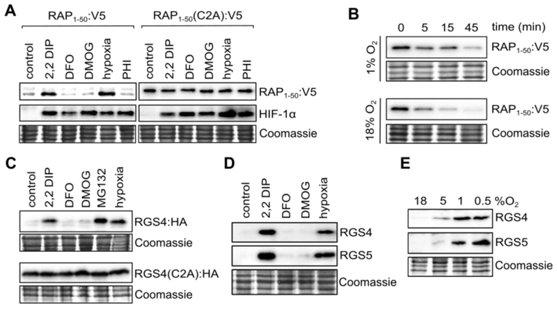
Organisms must respond to hypoxia to preserve oxygen homeostasis. We identify a thiol oxidase, previously assigned as cysteamine (2-aminoethanethiol) dioxygenase (ADO), as a low oxygen affinity (high-K mO2) amino-terminal cysteine dioxygenase that transduces the oxygen-regulated stability of proteins by the N-degron pathway in human cells. ADO catalyzes the conversion of amino-terminal cysteine to cysteine sulfinic acid and is related to the plant cysteine oxidases that mediate responses to hypoxia by an identical posttranslational modification. We show in human cells that ADO regulates RGS4/5 (regulator of G protein signaling) N-degron substrates, modulates G protein-coupled calcium ion signals and mitogen-activated protein kinase activity, and that its activity extends to other N-cysteine proteins including the angiogenic cytokine interleukin-32. Identification of a conserved enzymatic oxygen sensor in multicellular eukaryotes opens routes to better understanding and therapeutic targeting of adaptive responses to hypoxia.


Conflict of interest statement

Competing interests: PJR is a scientific co-founder holds equity in Reox Ltd, has served on the Research Advisory Board of GSK Ltd., and is co-inventor on patents disclosing methods and means of assaying for HIF hydroxylases inhibitors and their use in the treatment of hypoxic diseases (US8535899, EP1379630), which have been licensed to Reox Ltd. PJR, FL, EF, NM, TK, BG, MDW are inventors on a patent application related to this work (United Kingdom Patent Application No. 1908332.8). The other authors declare no competing interests.

Figures

Fig. 1
Fig. 1. Regulation of plant and animal N-degron substrates by oxygen in human cells.
(A) Levels of fusion proteins linking the N-terminal 1-50 residues of plant RAP2.12 or a C2A mutant to a GFP:V5 cassette (RAP1-50:V5; RAP1-50(C2A):V5) in stably transfected U-2OS cells exposed to hypoxia or the indicated inhibitors. (B) RAP1-50:V5 reporter protein half-life in cells incubated in hypoxia (16 h, 1% O2) then treated with cycloheximide (100 μM, 10min), then maintained in hypoxia or re-oxygenated for the indicated times. (C) C-terminal hemagglutinin (HA) tagged human RGS4, (RGS4:HA) or a C2A mutant in stably transfected RKO cells exposed to hypoxia or inhibitors. (D and E) Endogenous RGS4 and RGS5 proteins in SH-SY5Y cells exposed to inhibitors (D) or graded hypoxia (E). Similar patterns of response were observed for the plant fusion-protein reporter, transfected RGS4:HA and endogenous RGS4/5 proteins; responses of exogenous proteins were abolished by C2A mutation. 2,2 DIP, 2,2-dipyridyl (100 μM); DFO, desferrioxamine (100 μM); DMOG, dimethyloxalylglycine (1 mM); PHI, prolyl hydroxylase inhibitor (125 μM); MG132, proteasomal inhibitor (25 μM). All exposures of cells to hypoxia or inhibitors were for 4 h unless otherwise indicated. In panel A HIF-1α immunoblots are provided for comparison.
Fig. 2
Fig. 2. ADO controls the oxygen dependent Cys-branch of the N-degron pathway.
(A) RGS4:HA and RGS4(C2A):HA protein levels in 293T cells after co-expression with either control (EV), ADO, CDO1 or PCO1. Cells were exposed to hypoxia (0.5% O2, 16 h) or maintained in air. Comparable enzyme levels were confirmed by separate FLAG-immunoblotting. (B) Endogenous RGS4 and RGS5 proteins in ADO-deficient SH-SY5Y cells (ADO KO); RGS4 and RGS5 are constitutive and insensitive to iron chelators or hypoxia. (C) Over-expression of ADO does not repress constitutive stabilization of RGS5 in ATE1-deficient (ATE1 KO) cells. (D) Expression of human ADO restores wild type phenotype in 4pco A. thaliana mutants; 3pco mutants which did not manifest this phenotype were unaffected by ADO. (E) Box plots showing relative mRNA level of hypoxia-inducible genes in wild type and pco mutant plants that express ADO. ADO significantly reduced expression of the hypoxia-inducible genes PDC1, ADH, LBD41 and SAD6 in 4pco mutants, Mean ± S.D. *P<0.05; Mann-Whitney Rank Sum Test) with levels of non-hypoxia-inducible genes unchanged (fig. S10). (F) Relative luciferase activity (Fluc/Rluc) in S. cerevisiae cells expressing C-DLOR (Cys) or A-DLOR (Cys to Ala mutant) reporter under aerobic and hypoxic conditions in the presence or absence of human ADO or CDO1, mean ± S.D. *P<0.05; 2-way ANOVA followed by Holm-Sidak post hoc test.
Fig. 3
Fig. 3. ADO catalyzes the dioxygenation of the N-terminal Cys of RGS4/5 peptides.
(A) MS spectra show a mass shift of +32 Da when RGS5 N-terminal peptide was incubated with recombinant human ADO, but not with recombinant human CDO1. Similar results were obtained when an RGS4 N-terminal peptide was used (fig. S13). (B) The ADO-catalyzed +32 Da mass addition is absent when reactions were conducted under anaerobic (100% N2) conditions; 18O labelling demonstrates incorporation of 2 oxygen atoms derived directly from molecular O2 and not H2O. (C) Summary table of reaction kinetics for ADO-catalyzed dioxygenation of N-terminal RGS4/5 peptides. The influence of varying peptide concentration under atmospheric conditions (kcatPep and KmPepapp) and O2 levels using a fixed, non-limiting concentration of peptide (kcatO2 and KmO2app) were examined to determine sensitivities to both substrates. Source data are in fig. S15.
Fig. 4
Fig. 4. ADO regulates G-protein signalling.
(A-D) ADO regulates G-protein signalling in SH-SY5Y cells. (A) MAPK (p44/42) phosphorylation in WT, ADO KO and ATE1 KO cells. Immunoblot lanes represent separate biological replicates, with densitometric analysis provided below. Mean ± S.D. n=3, ***P<0.001 one-way ANOVA with Holm-Sidak post hoc test. (B) Re-expression of ADO increases phosphorylated p44/42 in ADO KO, but not ATE1 KO cells, mean ± S.D. n=3, **P<0.01 two-way ANOVA with Holm-Sidak post hoc test. (C) Carbachol (CCh) stimulated rises in [Ca2+]i are attenuated in ADO-deficient (KO) compared with wild-type (WT) cells. A representative trace is provided and mean peak change in R405/495 intensity at each CCh concentration is shown (inset). n=8-12, ***P<0.001, 3-parameter non-linear regression analysis. (D) Ionomycin (0.1μM) is equipotent at stimulating Ca2+ release in ADO KO cells infected with either control (EV) or ADO-containing lentivirus, whereas responses to CCh are recovered by ADO re-expression. Mean ± S.D. n=6-7, *P<0.05, two-way ANOVA with Holm-Sidak post hoc test.
Fig. 5
Fig. 5. IL-32 is a target of ADO-catalyzed N-terminal cysteine dioxygenation.
(A) Mass spectrometry analyses of the indicated IL-32 N-terminal peptide incubated aerobically with or without recombinant ADO (1h; 37°C), showing a +32 Da shift when incubated with ADO, indicative of the addition of O2. The small peak at ~845 m/z in the absence of ADO (top panel) was confirmed to correspond to a potassium adduct of the un-oxidized peptide. (B) IL-32, but not asparagine synthetase (ASNS) or JunB, are regulated by hypoxia (1% O2, 4 h) and ADO in RKO cells. (C) ADO-dependent regulation of IL-32 by hypoxia is observed at the protein but not mRNA level. (D) 293T cells co-transfected with plasmids encoding C-terminally FLAG-tagged IL-32 or an IL-32(C2A) mutant, and either empty pRRL vector (EV), ADO or a catalytically inactive ADO mutant (H112A+H114A), and exposed to hypoxia (1% O2) for 16 h. IL-32 levels were assessed using an anti-FLAG antibody. Hypoxic accumulation of IL-32 was evident in EV and mutant ADO, but not ADO, co-transfected cells, whilst C2A mutation abolished sensitivity to both hypoxia and ADO overexpression. Note that co-transfection with mutant ADO appears to increase basal levels of IL-32, consistent with possible competition with endogenous ADO for substrate binding.

Comment in

Similar articles

Cited by

References

    1. Epstein ACR, et al. C. elegans EGL-9 and mammalian homologues define a family of dioxygenases that regulate HIF by prolyl hydroxylation. Cell. 2001;107:43–54. - PubMed
    1. Ivan M, et al. HIFa targeted for VHL-mediated destruction by proline hydroxylation: implications for O2 sensing. Science. 2001;292:464–468. - PubMed
    1. Jaakkola P, et al. Targeting of HIF-a to the von Hippel-Lindau ubiquitylation complex by O2-regulated prolyl hydroxylation. Science. 2001;292:468–472. - PubMed
    1. Kaelin WGJ, Ratcliffe PJ. Oxygen sensing by metazoans: The central role of the HIF hydroxylase pathway. Molecular Cell. 2008;30:393–402. - PubMed
    1. West CM, van der Wel H, Wang ZA. Prolyl 4-hydroxylase-1 mediates O2 signaling during development of Dictyostelium. Development. 2007;134:3349–3358. - PubMed

Publication types