. 2019 Jul 4;9(1):9681.
doi: 10.1038/s41598-019-46094-y.

Human Corneal Expression of SLC4A11, a Gene Mutated in Endothelial Corneal Dystrophies

Affiliations

Human Corneal Expression of SLC4A11, a Gene Mutated in Endothelial Corneal Dystrophies

Darpan Malhotra et al. Sci Rep. .

Abstract

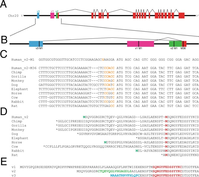
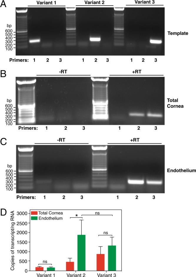
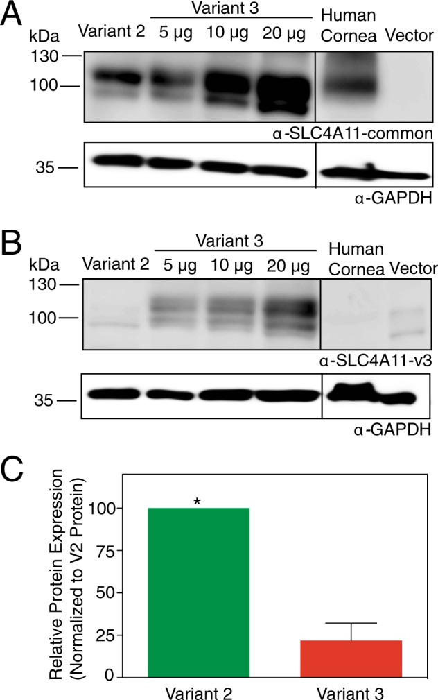
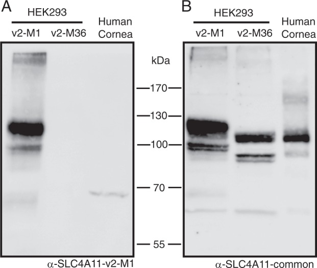
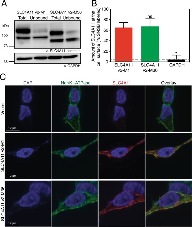
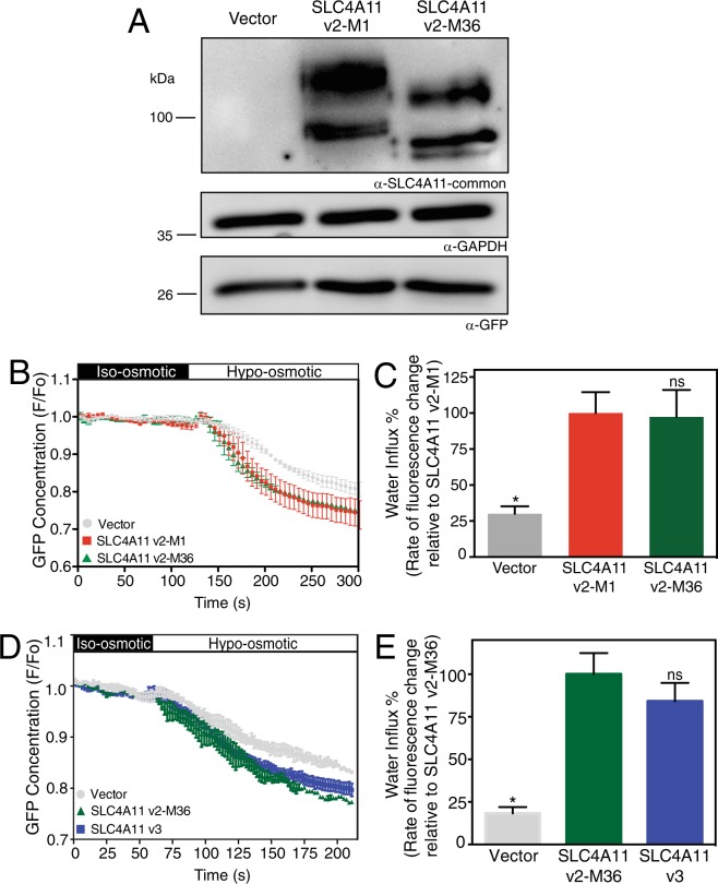
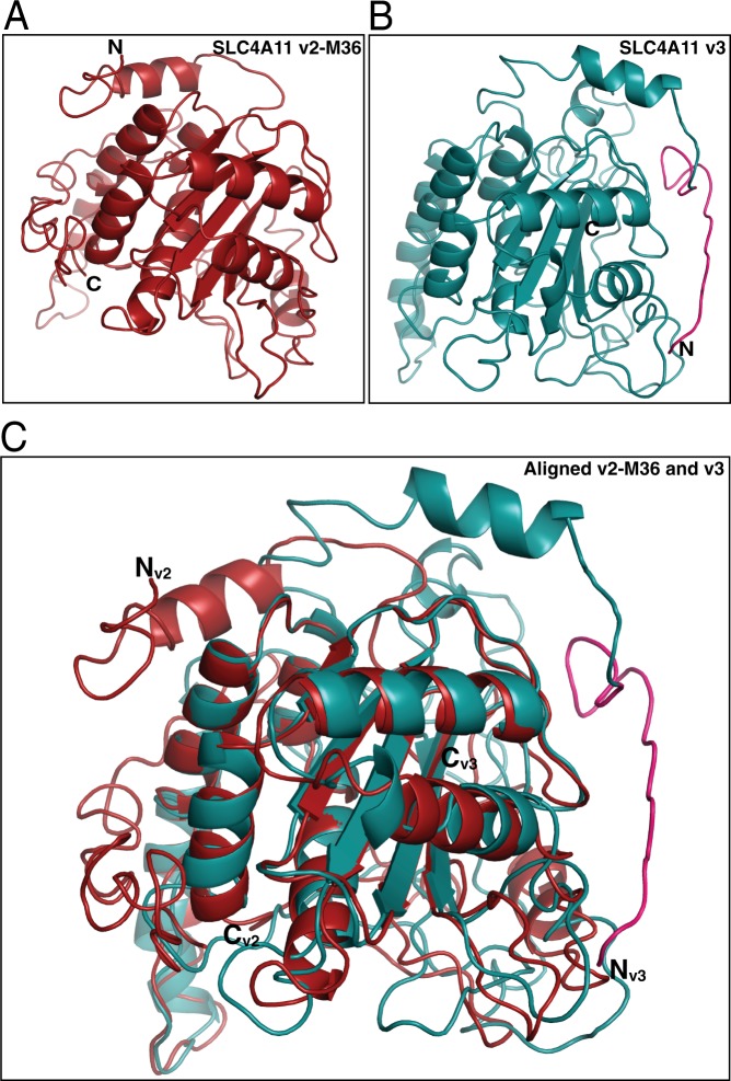
Two blinding corneal dystrophies, pediatric-onset congenital hereditary endothelial dystrophy (CHED) and some cases of late-onset Fuchs endothelial corneal dystrophy (FECD), are caused by SLC4A11 mutations. Three N-terminal SLC4A11 variants: v1, v2 and v3 are expressed in humans. We set out to determine which of these transcripts and what translated products, are present in corneal endothelium as these would be most relevant for CHED and FECD studies. Reverse transcription PCR (RT-PCR) and quantitative RT-PCR revealed only v2 and v3 mRNA in human cornea, but v2 was most abundant. Immunoblots probed with variant-specific antibodies revealed that v2 protein is about four times more abundant than v3 in human corneal endothelium. Bioinformatics and protein analysis using variant-specific antibodies revealed that second methionine in the open reading frame (M36) acts as translation initiation site on SLC4A11 v2 in human cornea. The v2 variants starting at M1 (v2-M1) and M36 (v2-M36) were indistinguishable in their cell surface trafficking and transport function (water flux). Structural homology models of v2-M36 and v3 suggest structural differences but their significance remains unclear. A combination of bioinformatics, RNA quantification and isoform-specific antibodies allows us to conclude that SLC4A11 variant 2 with start site M36 is predominant in corneal endothelium.


Conflict of interest statement

The authors declare no competing interests.

Figures

Figure 1
Figure 1
Bioinformatic analysis of human SLC4A11. (A) cDNA sequences of known human SLC4A11 transcripts were mapped onto a 15 kb segment of Human chromosome 20 genomic DNA to show transcript structure (coloured boxes), connected by introns (black lines). Each transcript variant (v1, v2 and v3) has a unique starting exon which results in unique N-terminal regions of the predicted protein and a common core region (red). (B) Magnified view of the 5′ end of the SLC4A11 gene. Each variant has a predicted start codon (M1; vertical black line). M36 (numbering based on predicted sequence of v2) marks the start of the common coding sequence for all transcripts. (C) Alignment of DNA sequences surrounding the predicted start codons in SLC4A11 v2 for multiple species. Kozak translational start sequence efficiency is indicated with bases matching the consensus shown in orange. For human SLC4A11, Kozak analysis is indicated for v2 start at Met 1 and 36 (v2-M1 and v2-M36). (D) Alignment of amino acid sequences for v2 SLC4A11 for the indicated mammals. Red Methionine (M) residue indicates the position corresponding to human v2-M36. Green Met indicates in frame Met upstream of v2-M36. Stop codons are indicated (*). (E) Amino acid sequences of SLC4A11 v1-v3 are aligned with sequences of peptides used to generate splice form-selective antibodies highlighted in red: SLC4A11-common, green: SLC4A11-v2-M1 and blue: SLC4A11-v3.
Figure 2
Figure 2
Expression of SLC4A11 transcripts in human cornea. (A) Plasmid-cloned SLC4A11 v1, v2 and v3 were each used as a template with specific forward primers for v1, v2 and v3, respectively and a common reverse primer. PCR reaction was set up for 40 cycles, and total reaction mixture was loaded on a 1% agarose gel. (B,C) RNA isolated from total cornea and micro-dissected endothelial layer were subjected to reverse transcription reaction with (+RT) or without (−RT) reverse transcriptase enzyme. PCR was performed using cDNA template generated from (B) total cornea and (C) isolated endothelium, with forward primers corresponding to v1, v2 and v3 along with common reverse primer. Expected amplicon sizes were 244, 233 and 233 bp for v1, v2 and v3, respectively. (D) qPCR was performed on cDNA from corneal endothelium and total cornea using iQ SYBR green mix along with forward and reverse primers for v1, v2 and v3 and GAPDH. Reactions were carried out for 40 cycles and fluorescence was recorded at each elongation step. Standard curve was constructed for each variant by using respective synthetic templates in the range of 2.5–2.5 × 104 copies and plotting Log10 (template copies) against their CT values. The sample CT values were corrected to GAPDH for every experiment and number of copies of each variant was calculated, using their respective standard curves. Background (-RT control) values were subtracted from the samples. The average number of copies per ng RNA were plotted for each variant in corneal endothelium and total cornea. Data are presented as mean ± SEM from three biological replicates (Two-way ANOVA and Sidak’s multiple comparison test). *p = 0.02, ns = not significant.
Figure 3
Figure 3
SLC4A11 v2 and v3 expression in human corneal endothelium. Corneal endothelium was micro-dissected from six human corneas. Protein lysates from pooled human corneal endothelium (20 µg protein) along with HEK293 cells transfected with cDNA encoding SLC4A11 v2 (10 µg protein lysate), v3 (5, 10, 20 µg protein lysates, indicated) and empty vector (20 µg protein lysate) were processed for immunoblots. Blots were probed with (A) SLC4A11-common antibody and (B) SLC4A11-v3 antibody, along with GAPDH antibody as a loading control. (C) Densitometry was performed to quantify band intensities. Detection ratio was established between SLC4A11-common antibody and SLC4A11-v3 antibody (see Supplementary Fig. S1). Data from each trial were normalized to SLC4A11 v2 abundance. Data represent mean ± SEM from three replicates (Two-tailed t-test). *p = 0.005 when compared to v3. Note that intervening lanes to the left of the human cornea lane in (A) and (B) have been removed and are marked by the black line.
Figure 4
Figure 4
Identification of SLC4A11 v2 translation start site in total human corneal lysates. HEK293 cells were transfected with cDNA encoding: SLC4A11 v2 starting at Met1 (SLC4A11 v2-M1) or SLC4A11 v2 starting at Met36 (SLC4A11 v2-M36). Whole human cornea samples were flash frozen with liquid nitrogen, crushed, solubilized and then combined with 2X SDS-Page sample buffer. Samples were electrophoresed on 7.5% acrylamide gels. Corresponding immunoblots were probed with antibodies (A) Anti-SLC4A11-v2-M1 and (B) Anti-SLC4A11-common.
Figure 5
Figure 5
Plasma membrane localization of SLC4A11 v2-M1 and v2-M36. HEK293 cells were transfected with cDNA encoding SLC4A11 v2-M1 or SLC4A11 v2-M36 as shown. (A) Cells were labeled with membrane-impermeant Sulfo-NHS-SS-biotin (SNSB) and the cell lysates were divided into two equal fractions. One was incubated with streptavidin resin to remove biotinylated proteins. The unbound fraction (U) and the total cell lysate (T) were probed with SLC4A11-common antibody on immunoblots. GAPDH was used as an internal control. (B) The fraction of SLC4A11 and GAPDH labeled by SNSB was calculated. Dashed line indicates the background for this assay, as indicated by labeling of GAPDH. Data represent mean ± SEM from five replicates (One-way ANOVA with Sidak’s multiple comparisons test). *p < 0.0001 when compared to SLC4A11 v2-M1, ns = not significant. (C) Cells were processed for immunofluorescence and images were collected by confocal microscopy (scale bars indicated in each row). Nuclei were detected with DAPI staining (blue). SLC4A11 was detected with SLC4A11-common antibody and chicken anti-rabbit IgG conjugated with Alexa Fluor 594 (red). Plasma membrane marker, Na+/K+-ATPase was detected with anti-Na+/K+-ATPase and chicken anti-mouse Alexa Fluor 488 (green). Overlay represents merged images from all three channels.
Figure 6
Figure 6
Osmotically driven water flux activity of SLC4A11 v2-M1 and v2-M36 and v3. HEK293 cells were transiently transfected with cDNA encoding eGFP along with empty vector or SLC4A11 v2-M1 or SLC4A11 v2-M36. (A) After performing water flux assays, cells from the same dishes were lysed and probed on immunoblots, using SLC4A11-common antibody to detect the expression of SLC4A11 variants. GAPDH and eGFP were also detected using respective antibodies. (B,D) HEK293 cells transfected with v2-M1 or v2-M36 or v3 cDNA were perfused alternately with iso-osmotic (black bar) and hypo-osmotic (white bar) media. eGFP fluorescence (F) was normalized to F averaged for time 0–120 or 0–60 seconds (F0) and F/F0 plotted. (C,E) Rate of fluorescence change (a surrogate for cell volume change) was calculated. Data represent mean ± SEM from 4–8 independent coverslips with 12–18 cells measured per coverslip (One-way ANOVA with Sidak’s multiple comparisons test) *p < 0.0001 when compared to SLC4A11 v2-M1 or SLC4A11 v2-M36, ns = not significant.
Figure 7
Figure 7
Homology model of SLC4A11 v2-M36 and SLC4A11 v3 cytoplasmic domains. Homology models of cytoplasmic domains of (A) SLC4A11 v2-M36 (M36-Y340) and (B) SLC4A11 v3 (M1-Y359) generated by I-TASSER using crystal structure of human erythrocyte Band 3 cytoplasmic domain (PDB: 1HYN) as the template. Unique N-terminal sequence of v3 is highlighted in pink. (C) v2-M36 and v3 models aligned in PyMOL (molecular graphics system version 2.0.7), using segments that were modelled with 100% confidence (R90-P306 for v2-M36, R109-P325 for v3). Nv2, Nv3 and Cv2, Cv3 are the N and C termini for v2-M36 and v3, respectively.

Similar articles

Cited by

References

    1. Klintworth GK. Corneal dystrophies. Orphanet. J. Rare Dis. 2009;4:7. doi: 10.1186/1750-1172-4-7. - DOI - PMC - PubMed
    1. Bonanno JA. Identity and regulation of ion transport mechanisms in the corneal endothelium. Prog Retin Eye Res. 2003;22:69–94. doi: 10.1016/S1350-9462(02)00059-9. - DOI - PubMed
    1. 2016 Eye Banking Statisitical Report. (Eye Bank Association of America, Washington, D.C., U.S.A., 2017).
    1. Vithana EN, et al. SLC4A11 Mutations in Fuchs Endothelial Corneal Dystrophy (FECD) Hum. Mol. Genet. 2008;17:656–666. doi: 10.1093/hmg/ddm337. - DOI - PubMed
    1. Biswas S, et al. Missense mutations in COL8A2, the gene encoding the alpha2 chain of type VIII collagen, cause two forms of corneal endothelial dystrophy. Hum. Mol. Genet. 2001;10:2415–2423. doi: 10.1093/hmg/10.21.2415. - DOI - PubMed

Publication types