

Morphological Brain Age Prediction using Multi-View Brain Networks Derived from Cortical Morphology in Healthy and Disordered Participants
- PMID: 31273275
- PMCID: PMC6609705
- DOI: 10.1038/s41598-019-46145-4
Morphological Brain Age Prediction using Multi-View Brain Networks Derived from Cortical Morphology in Healthy and Disordered Participants
Abstract
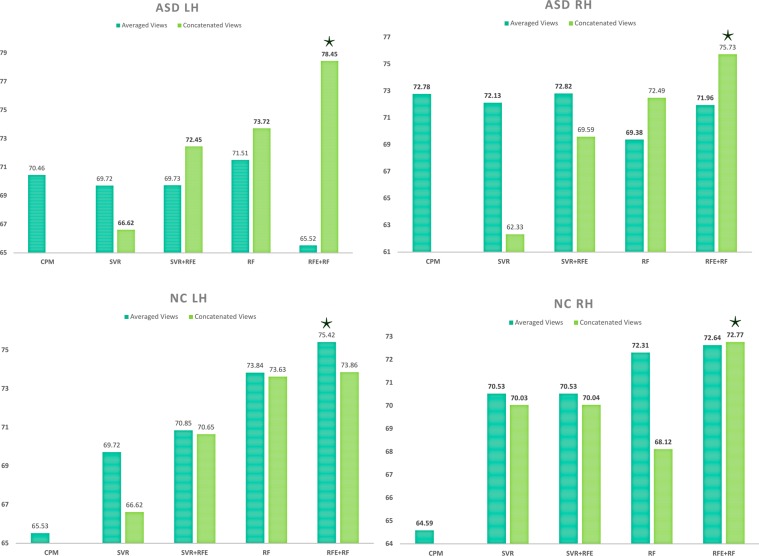
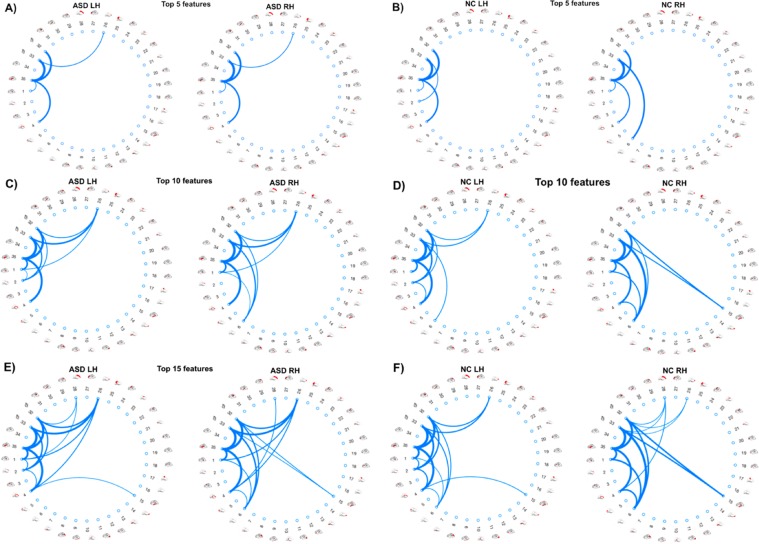
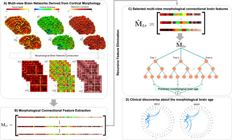
Brain development and aging are dynamic processes that unfold over years on multiple levels in both healthy and disordered individuals. Recent studies have revealed a disparity between the chronological brain age and the 'data-driven' brain age using functional MRI (fMRI) and diffusion MRI (dMRI). Particularly, predicting the 'brain age' from connectomic data might help identify relevant connectional biomarkers of neurological disorders that emerge early or late in the lifespan. While prior brain-age prediction studies have relied exclusively on either structural or functional connectomic data, here we unprecedentedly propose to predict the morphological age of the brain by solely using morphological brain networks (derived from T1-weighted images) in both healthy and disordered populations. Besides, although T1-weighted MRI was widely used for brain age prediction, it was leveraged from an image-based analysis perspective not from a connectomic perspective. Our method includes the following steps: (i) building multi-view morphological brain networks (M-MBN), (ii) feature extraction and selection, (iii) training a machine-learning regression model to predict age from M-MBN data, and (iv) utilizing our model to identify connectional brain features related to age in both autistic and healthy populations. We demonstrate that our method significantly outperforms existing approaches and discovered brain connectional morphological features that fingerprint the age of brain cortical morphology in both autistic and healthy individuals. In particular, we discovered that the connectional cortical thickness best predicts the morphological age of the autistic brain.
Conflict of interest statement
The authors declare no competing interests.
Figures
Similar articles
-
Joint functional brain network atlas estimation and feature selection for neurological disorder diagnosis with application to autism.Med Image Anal. 2020 Feb;60:101596. doi: 10.1016/j.media.2019.101596. Epub 2019 Nov 7. Med Image Anal. 2020. PMID: 31739282
-
Clustering-based multi-view network fusion for estimating brain network atlases of healthy and disordered populations.J Neurosci Methods. 2019 Jan 1;311:426-435. doi: 10.1016/j.jneumeth.2018.09.028. Epub 2018 Sep 30. J Neurosci Methods. 2019. PMID: 30282004
-
Estimation of gender-specific connectional brain templates using joint multi-view cortical morphological network integration.Brain Imaging Behav. 2021 Aug;15(4):2081-2100. doi: 10.1007/s11682-020-00404-5. Epub 2020 Oct 21. Brain Imaging Behav. 2021. PMID: 33089469 Free PMC article.
-
A systematic review of structural MRI biomarkers in autism spectrum disorder: A machine learning perspective.Int J Dev Neurosci. 2018 Dec;71:68-82. doi: 10.1016/j.ijdevneu.2018.08.010. Epub 2018 Aug 30. Int J Dev Neurosci. 2018. PMID: 30172895
-
Spatio-temporal modeling of connectome-scale brain network interactions via time-evolving graphs.Neuroimage. 2018 Oct 15;180(Pt B):350-369. doi: 10.1016/j.neuroimage.2017.10.067. Epub 2017 Nov 10. Neuroimage. 2018. PMID: 29102809 Free PMC article. Review.
Cited by
-
Cognitive impairment in adolescent and young adult cancer patients: Pre-treatment findings of a longitudinal study.Cancer Med. 2023 Feb;12(4):4821-4831. doi: 10.1002/cam4.5295. Epub 2022 Oct 11. Cancer Med. 2023. PMID: 36221816 Free PMC article.
-
Brain-age estimation accuracy is significantly increased using multishell free-water reconstruction.Hum Brain Mapp. 2022 May;43(7):2365-2376. doi: 10.1002/hbm.25792. Epub 2022 Feb 10. Hum Brain Mapp. 2022. PMID: 35141974 Free PMC article.
-
In patients with mild disability NMOSD: is the alteration in the cortical morphological or functional network topological properties more significant.Front Immunol. 2024 Feb 5;15:1345843. doi: 10.3389/fimmu.2024.1345843. eCollection 2024. Front Immunol. 2024. PMID: 38375481 Free PMC article.
-
Explainable Deep Learning for Personalized Age Prediction With Brain Morphology.Front Neurosci. 2021 May 28;15:674055. doi: 10.3389/fnins.2021.674055. eCollection 2021. Front Neurosci. 2021. PMID: 34122000 Free PMC article.
-
Predicting 'Brainage' in the Developmental Period using Structural MRI, Morphometric Similarity, and Machine Learning.Res Sq [Preprint]. 2023 Feb 28:rs.3.rs-2583936. doi: 10.21203/rs.3.rs-2583936/v1. Res Sq. 2023. Update in: Sci Rep. 2023 Sep 20;13(1):15591. doi: 10.1038/s41598-023-42414-5. PMID: 36909598 Free PMC article. Updated. Preprint.
References
Publication types
MeSH terms
LinkOut - more resources
Full Text Sources
Other Literature Sources
Medical
Miscellaneous
