Review
. 2019 Sep;46(10):1990-2012.
doi: 10.1007/s00259-019-04404-6. Epub 2019 Jul 4.

Two decades of SPECT/CT - the coming of age of a technology: An updated review of literature evidence

Affiliations
Review

Two decades of SPECT/CT - the coming of age of a technology: An updated review of literature evidence

Ora Israel et al. Eur J Nucl Med Mol Imaging. 2019 Sep.

Abstract

Purpose: Single-photon emission computed tomography (SPECT) combined with computed tomography (CT) was introduced as a hybrid SPECT/CT imaging modality two decades ago. The main advantage of SPECT/CT is the increased specificity achieved through a more precise localization and characterization of functional findings. The improved diagnostic accuracy is also associated with greater diagnostic confidence and better inter-specialty communication.

Methods: This review presents a critical assessment of the relevant literature published so far on the role of SPECT/CT in a variety of clinical conditions. It also includes an update on the established evidence demonstrating both the advantages and limitations of this modality.

Conclusions: For the majority of applications, SPECT/CT should be a routine imaging technique, fully integrated into the clinical decision-making process, including oncology, endocrinology, orthopaedics, paediatrics, and cardiology. Large-scale prospective studies are lacking, however, on the use of SPECT/CT in certain clinical domains such as neurology and lung disorders. The review also presents data on the complementary role of SPECT/CT with other imaging modalities and a comparative analysis, where available.

Keywords: Cardiopulmonary; Endocrinology; Infection; Oncology; Orthopaedics; Paediatrics; SPECT/CT.


Conflict of interest statement

Ora Israel: consultant for GE Healthcare

Gopinath Gnanasegaran: symposia attendance support, Norgine Radiopharmaceuticals

Torsten Kuwert: speaker honoraria, Siemens Healthineers and Sanofi; institutional research grant, Siemens Healthineers; institutional material support, Progenics

Christian la Fougère: consultant, speaker honoraria, research grant, GE Healthcare; research grants, Siemens Healthineers; consultant for Bayer

Samia Massalha: Tucker Research Fellowship award, University of Ottawa Heart Institute

Olivier Pellet, Lorenzo Biassoni, Diego De Palma, Enrique Estrada-Lobato, Giuliano Mariani, Diana Paez D and Francesco Giammarile have no conflicts of interest to declare

Figures

Fig. 1
Fig. 1
99mTc-nanocolloid SPECT/CT in sentinel node lymphoscintigraphy. Preoperative sentinel lymph node (SLN) mapping in a 63-year-old woman with cancer of the left breast following intratumoral injection of 99mTc-nanocolloidal albumin. Planar imaging with body contouring (a), with a 57Co flood source beneath the patient’s body, visualizes lymphatic drainage to SLNs in the left axilla, periclavicular area, and internal mammary chain. 3D surface volume rendering SPECT/CT (b) identifies the anatomical correlates of the SLNs localized on transaxial SPECT/CT and CT slices to the second intercostal space and level I of the left axilla, respectively (c, c), as well as behind the left clavicle (e, f), as indicated by dashed yellow circles. [Reproduced with permission from: Giammarile F, Orsini F, Valdés Olmos RA, Vidal-Sicart S, Giuliano AE, Mariani G. Radioguided surgery for breast cancer. In: Strauss HW, Mariani G, Volterrani D, Larson SM, Eds. Nuclear Oncology – From Pathophysiology to Clinical Applications. New York: Springer; 2017:1363–1400]
Fig. 2
Fig. 2
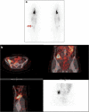
Post-ablation 131I-iodide SPECT/CT in differentiated thyroid cancer (DTC). A 48-year-old woman after thyroidectomy and lymphadenectomy for DTC (18 mm, infiltrating the thyroid parenchyma and capsule), with intravascular tumour emboli and nodal metastases found at surgery, stage pT1bN1bMx. The patient received 2.96 GBq 131I-iodide treatment. Planar post-ablation 131I-iodide scan (a) shows multiple sites of intense uptake in the neck and upper mediastinum consistent with known metastatic spread. There is an additional area of focal tracer accumulation of moderate intensity in the right pelvis (arrow). SPECT/low-dose CT slices and maximum intensity projection (MIP) image of the pelvis (b) locate this uptake to the lower portion of the ascending colon, consistent with physiologic tracer excretion. SPECT/CT excluded the presence of an additional malignant site in an equivocal finding detected on planar scintigraphy. [Images provided courtesy of Drs. Paola A. Erba and Roberta Zanca, Regional Center of Nuclear Medicine, University of Pisa, Italy]
Fig. 3
Fig. 3
111In-somatostatin SPECT/CT in neuroendocrine neoplasms. A 34-year-old patient with biochemical suspicion of NEN and a lesion in the head of pancreas on CT was referred for 111In-somatostatin scintigraphy for staging. SPECT (a) shows two foci of intense tracer uptake in the upper abdomen, localized by contrast-enhanced CT (b) to a hypervascular primary lesion in the head of the pancreas and an isodense metastasis in the right lobe of the liver (arrows), confirmed on SPECT/CT (c)
Fig. 4
Fig. 4
99mTc-DPD bone SPECT/CT in a patient with renal cancer and bone pain. A 62-year-old patient with renal cancer was referred for evaluation of painful left elbow. Early planar scan of the elbows (a) shows intense hyperaemia on the left side. Delayed whole-body scintigraphy (b, posterior view) demonstrates intense inhomogeneous focal uptake in the distal part of the left humerus, localized by SPECT/CT (c) to an osteolytic metastasis seen on the CT component
Fig. 5
Fig. 5
99mTc-MIP-1404 PSMA-SPECT/CT in a patient with prostate cancer. An 81-year-old man with prostate cancer (pT4, N2, M0, G3) with biochemical recurrence diagnosed by increased serum PSA levels from 6.59 ng/mL to 17.34 ng/mL over a 3-month interval, was referred for restaging. Whole-body SPECT-MIP (a) shows multiple sites of focal abnormal tracer uptake above and below the diaphragm. Transaxial SPECT/CT and CT slices at the level of the upper thorax (b) demonstrate the presence of a left internal mammary chain lymph node metastasis. Transaxial SPECT/CT and CT slices at the level of the upper pelvis (c) show multiple, partially MIP-1404-avid osteoblastic lesions in the bone, consistent with functionally active and inactive skeletal metastases
Fig. 6
Fig. 6
99mTc-MDP bone SPECT/CT in a patient with a painful left foot. A 70-year-old patient with persistent pain in the left foot was referred for bone scintigraphy (BS) for detection of the pain generator. Early planar scan of the feet (a) and delayed whole-body scan (b) show a focus of hyperaemia and intense tracer uptake in the posterior aspect of the left tarsal region, localized by SPECT/CT (c) to severe degenerative changes in the left talonavicular joint seen on the CT component
Fig. 7
Fig. 7
99mTc-HMPAO-labelled leucocyte SPECT/CT in a patient with diabetic foot. A 43-year-old man with diabetes mellitus and an infected wound in the medial aspect of the left forefoot was referred for 99mTc-HMPAO-leucocyte scintigraphy for suspected osteomyelitis. Planar scans (a) performed at 1 h (top), 4 h (center), and 24 h (bottom) after tracer injection show a focal area of uptake at the base of the first left digit, increasing in intensity in its superior aspect. SPECT/CT (b) locates this uptake to fragments of the first left metatarsal bone showing areas of cortical erosion and sclerosis, as well as to the adjacent deep wound and surrounding edematous soft tissue, consistent with osteomyelitis in addition to the soft tissue infection
Fig. 8
Fig. 8
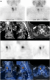
99mTc-Sestamibi SPECT/CT for localization of parathyroid adenoma (PTA). A 57-year-old woman with laboratory evidence of primary hyperparathyroidism was referred for localization of PTA. Planar scintigraphy (a) shows an early area of increased focal uptake at the upper pole of the right thyroid lobe (left), with washout of the tracer from adjacent thyroid tissue on late images (right). SPECT/CT (b) performed 1 h after tracer injection localizes this focal uptake to a 16-mm nodule behind the thyroid gland, consistent with a PTA
Fig. 9
Fig. 9
Lung SPECT/CT, perfusion study in suspected pulmonary embolism (PE). A 60-year-old patient with clinical suspicion of pulmonary embolism was referred for pulmonary ventilation/perfusion scintigraphy. Planar scintigraphy selected posterior and posterior oblique views) shows normal ventilation (a), in the presence of multiple segmental and subsegmental perfusion defects in both lungs (b). SPECT/CT shows no corresponding abnormal parenchymal changes on the CT component of the study (c). The patient was diagnosed with a bilateral pulmonary embolism
Fig. 10
Fig. 10
99mTc-tetrofosmin cardiac SPECT/CT for attenuation correction. A patient presenting with chest pain and with family history of CAD was referred for a myocardial perfusion SPECT/CT using a single-tracer one-day stress–rest protocol with 99mTc-tetrofosmin. CT AC was performed for the stress study. MPI-SPECT short (a), horizontal (b), and vertical (c) long axis show moderate reduction in tracer uptake in the anteroseptal wall sparing the anterobasal segment, and in the inferior wall and the apex on the non-corrected images at stress (first row), normalizing at rest (third row), suggests the presence of moderate ischemia in the territories of the left anterior descending (LAD) and right coronary (RCA) arteries. AC stress images (second row) confirm the presence of decreased uptake in the territory of the LAD only (the anteroseptal wall and apex), while correcting the attenuation artefact caused by the diaphragm in the inferior wall. The findings are consistent with a large area of moderate ischemia in the territory supplied by the LAD. Coronary angiography (c) demonstrates normal RCA (top, arrow) and a critical lesion in the proximal LAD (bottom, arrow)
Fig. 11
Fig. 11
123I-FP-CIT SPECT/CT in a patient with parkinsonism. A 63-year-old man with parkinsonian symptoms for the past 12 months, initially diagnosed as Parkinson’s disease but with no appreciable response to L-DOPA treatment, was referred to brain scintigraphy. Transaxial SPECT and SPECT/CT (centre and bottom row) show reduced tracer uptake in the left putamen. CT (top row) demonstrates ipsilateral cortical atrophy. These combined findings suggest a diagnosis of corticobasal atrophy with associated parkinsonism, rather than typical Parkinson’s disease. [Images provided courtesy of Dr. Duccio Volterrani, Regional Center of Nuclear Medicine, University of Pisa, Italy]
Fig. 12
Fig. 12
Bone SPECT/CT diagnosis of spondylolysis in an adolescent patient. A14-year-old girl was referred for evaluation of persistent low back pain. Early planar scan of lumbar spine and pelvis (a) shows no hyperaemia. Delayed WB-BS, anterior and posterior views (b) show a focus of abnormal 99mTc-MDP uptake in left aspect of L5 vertebra, localized by SPECT/CT (c) to the left L-5 articular facet, consistent with spondylolysis
Fig. 13
Fig. 13
123I-mIBG SPECT/CT in neuroblastoma. A 6-year-old child with newly diagnosed neuroblastoma was referred for staging. Planar WB scan (a) shows an area of increased uptake in the right upper abdomen, partially overlapping the liver of unclear location and clinical significance. SPECT/CT (b) localizes this abnormal tracer uptake to the primary lesion in a large mass in the right adrenal seen on CT (C)

References

    1. Alavi A, Basu S. Planar and SPECT imaging in the era of PET and PET–CT: can it survive the test of time? Eur J Nucl Med Mol Imaging. 2008;35:1554. - PubMed
    1. Mariani G, Strauss HW. Positron emission and single-photon emission imaging: synergy rather than competition. Eur J Nucl Med Mol Imaging. 2011;38:1189–1190. - PubMed
    1. Bischof Delaloye A, Carrió I, Cuocolo A, Knapp W, Gourtsoyiannis N, McCall I, et al. White paper of the European Association of Nuclear Medicine (EANM) and the European Society of Radiology (ESR) on multimodality imaging. Eur J Nucl Med Mol Imaging. 2007;34:1147–1151. - PubMed
    1. Kashyap R, Dondi M, Paez D, Mariani G, editors. Hybrid imaging worldwide—challenges and opportunities for the developing world: a report of a technical meeting organized by IAEA. Semin Nucl Med. 2013;43:208–23. - PubMed
    1. Mariani G, Bruselli L, Kuwert T, Kim EE, Flotats A, Israel O, et al. A review on the clinical uses of SPECT/CT. Eur J Nucl Med Mol Imaging. 2010;37:1959–1985. - PubMed

MeSH terms