

Virus-induced gene silencing (VIGS) in Lilium leichtlinii using the Cucumber mosaic virus vector
- PMID: 31274998
- PMCID: PMC6587034
- DOI: 10.5511/plantbiotechnology.16.1018a
Virus-induced gene silencing (VIGS) in Lilium leichtlinii using the Cucumber mosaic virus vector
Abstract
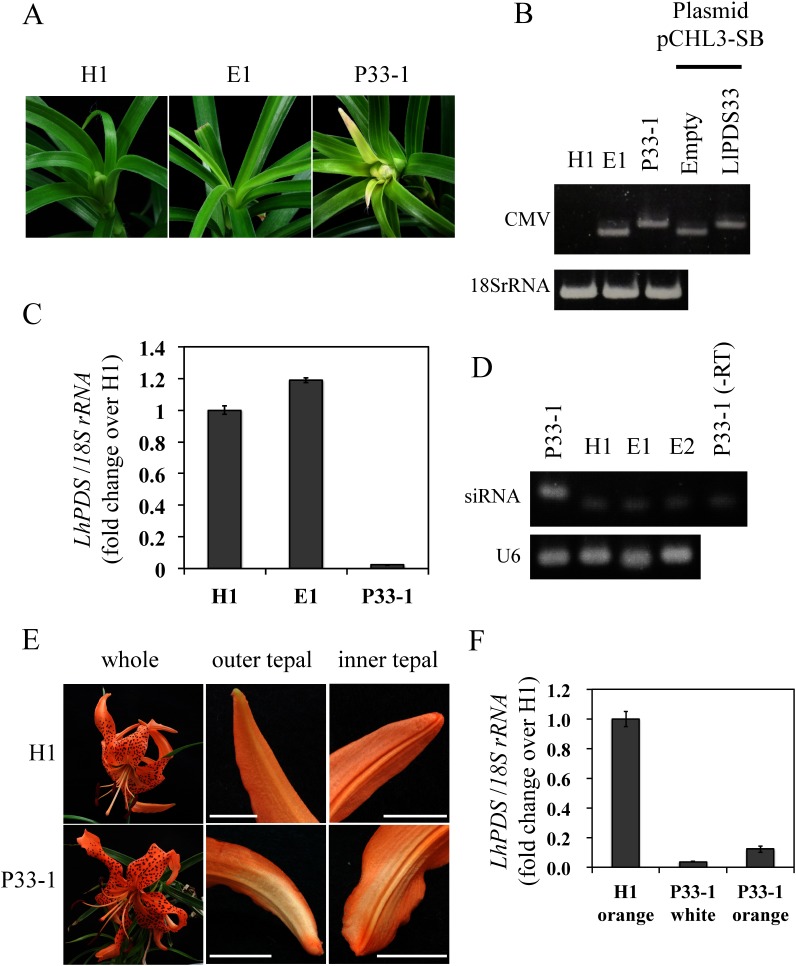
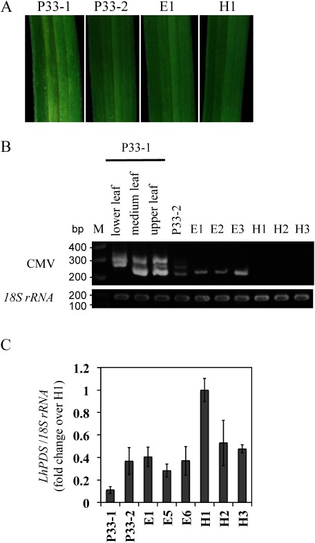
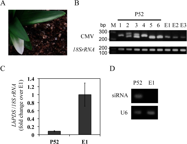
Lilies (Lilium) are among the most important floriculture crops, and to accelerate research regarding lily genetics, the development of reverse-genetics tools is necessary. However, Agrobacterium-mediated transformation in Lilium is time-consuming, since the plants require several years to progress from acclimation to flowering. Thus, virus-induced gene silencing (VIGS) is an attractive method for assaying gene function. In the present study, we modified a lily-derived strain of Cucumber mosaic virus (CMV-HL) as a VIGS vector and evaluated its effectiveness for inducing gene silencing in Lilium leichtlinii by introducing L. leichtlinii phytoene desaturase (LlPDS) gene fragments into an intercistronic region between the 3a and 3b genes of the CMV-HL RNA3 genome. At 30 days after inoculation (dpi) with LlPDS-containing CMV-HL, photo-bleaching was observed in the upper leaves of L. leichtlinii, and at 57 dpi, we observed that the natural orange color in flower tepals had faded. Reduced LlPDS expression and the detection of small interfering LlPDS RNA indicated that the color changes were the result of LlPDS gene silencing. In addition, the leaves also exhibited a mild photo-bleaching phenotype in the following year. Therefore, our results indicate that CMV-HL spreads systemically in the leaves and flowers of Lilium during the first year of infection, as well as in new shoots during the following year, and that the vector system can be successfully applied to induce short-term endogenous gene silencing in lilies.
Keywords: Cucumber mosaic virus (CMV)-HL; lily; photo-bleaching; phytoene desaturase (PDS); small interfering RNA (siRNA).
Figures
Similar articles
-
Application of cucumber mosaic virus to efficient induction and long-term maintenance of virus-induced gene silencing in spinach.Plant Biotechnol (Tokyo). 2020 Mar 25;37(1):83-88. doi: 10.5511/plantbiotechnology.19.1227a. Plant Biotechnol (Tokyo). 2020. PMID: 32362752 Free PMC article.
-
Virus-Induced Gene Silencing in Lilies Using Cucumber Mosaic Virus Vectors.Methods Mol Biol. 2020;2172:1-13. doi: 10.1007/978-1-0716-0751-0_1. Methods Mol Biol. 2020. PMID: 32557357
-
Establishment of virus-induced gene silencing (VIGS) system in Luffa acutangula using Phytoene desaturase (PDS) and tendril synthesis related gene (TEN).Plant Methods. 2023 Aug 31;19(1):94. doi: 10.1186/s13007-023-01064-4. Plant Methods. 2023. PMID: 37653449 Free PMC article.
-
Efficient and high-throughput pseudorecombinant-chimeric Cucumber mosaic virus-based VIGS in maize.Plant Physiol. 2021 Dec 4;187(4):2865-2876. doi: 10.1093/plphys/kiab443. Plant Physiol. 2021. PMID: 34606612 Free PMC article.
-
Development, progress and future prospects in cryobiotechnology of Lilium spp.Plant Methods. 2019 Nov 2;15:125. doi: 10.1186/s13007-019-0506-9. eCollection 2019. Plant Methods. 2019. PMID: 31700526 Free PMC article. Review.
Cited by
-
Advanced Biotechnological Interventions in Mitigating Drought Stress in Plants.Plants (Basel). 2024 Mar 4;13(5):717. doi: 10.3390/plants13050717. Plants (Basel). 2024. PMID: 38475564 Free PMC article. Review.
-
Horticultural innovation by viral-induced gene regulation of carotenogenesis.Hortic Res. 2022 Jan 18;9:uhab008. doi: 10.1093/hr/uhab008. Online ahead of print. Hortic Res. 2022. PMID: 35043183 Free PMC article.
-
Application of cucumber mosaic virus to efficient induction and long-term maintenance of virus-induced gene silencing in spinach.Plant Biotechnol (Tokyo). 2020 Mar 25;37(1):83-88. doi: 10.5511/plantbiotechnology.19.1227a. Plant Biotechnol (Tokyo). 2020. PMID: 32362752 Free PMC article.
-
Transcriptional silencing of 35S driven-transgene is differentially determined depending on promoter methylation heterogeneity at specific cytosines in both plus- and minus-sense strands.BMC Plant Biol. 2019 Jan 14;19(1):24. doi: 10.1186/s12870-019-1628-y. BMC Plant Biol. 2019. PMID: 30642254 Free PMC article.
-
Tobacco rattle virus-induced PHYTOENE DESATURASE (PDS) and Mg-chelatase H subunit (ChlH) gene silencing in Solanum pseudocapsicum L.PeerJ. 2018 Mar 20;6:e4424. doi: 10.7717/peerj.4424. eCollection 2018. PeerJ. 2018. PMID: 29576941 Free PMC article.
References
-
- Azadi P, Chin DP, Kuroda K, Khan RS, Mii M (2010) Macro elements in inoculation and co-cultivation medium strongly affect the efficiency of Agrobacterium-mediated transformation in Lilium. Plant Cell Tissue Organ Cult 101: 201–209
-
- Goto K, Kanazawa A, Kusaba M, Masuta C (2003) A simple and rapid method to detect plant siRNAs using nonradioactive probes. Plant Mol Biol Report 21: 51–58
-
- Hagita T, Kodama F, Akai J (1989) The virus diseases of lily in Hokkaido. Ann Phytopathological Soc Jpn 55: 1–8 (in Japanese)
LinkOut - more resources
Full Text Sources
Miscellaneous