. 2019 Jun 7:10:1267.
doi: 10.3389/fmicb.2019.01267. eCollection 2019.

Nanoscale Tungsten-Microbial Interface of the Metal Immobilizing Thermoacidophilic Archaeon Metallosphaera sedula Cultivated With Tungsten Polyoxometalate

Affiliations

Nanoscale Tungsten-Microbial Interface of the Metal Immobilizing Thermoacidophilic Archaeon Metallosphaera sedula Cultivated With Tungsten Polyoxometalate

Tetyana Milojevic et al. Front Microbiol. .

Abstract

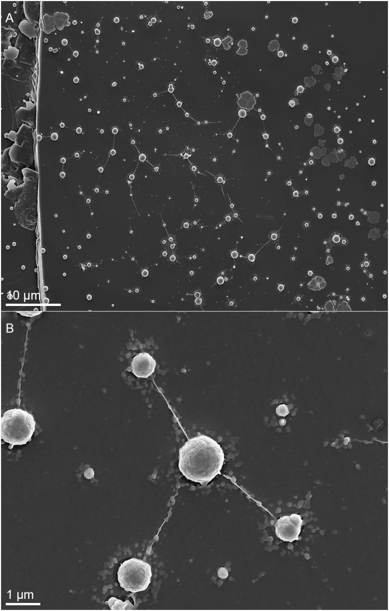
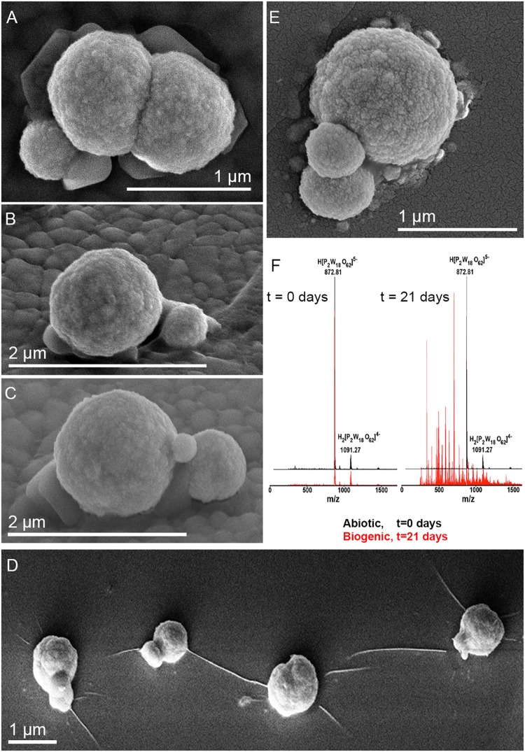
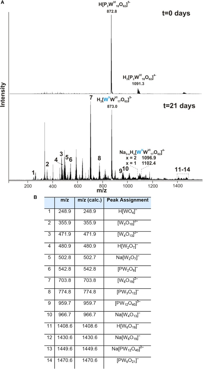
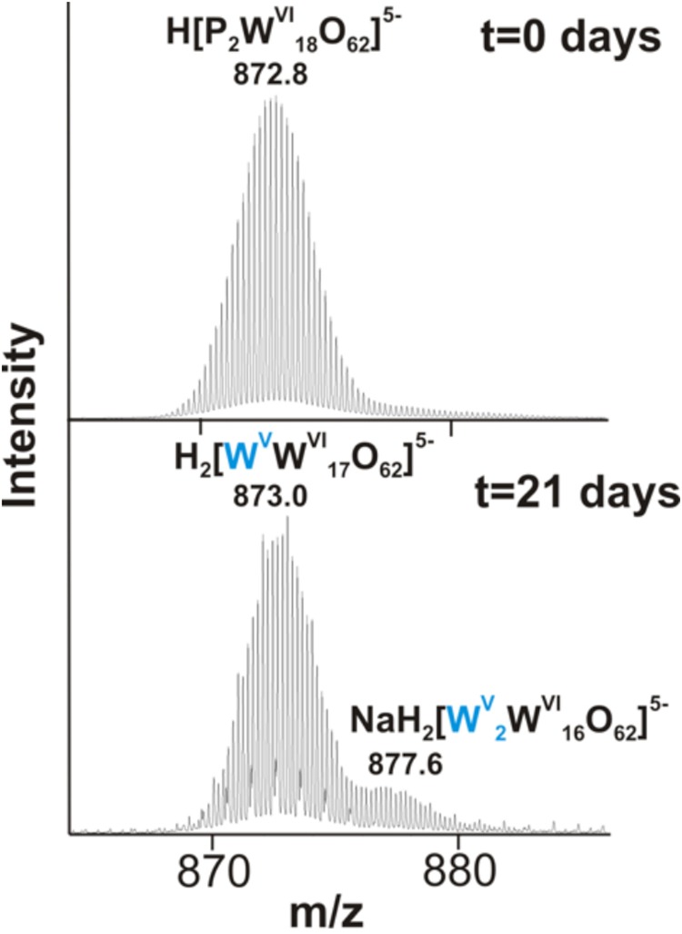
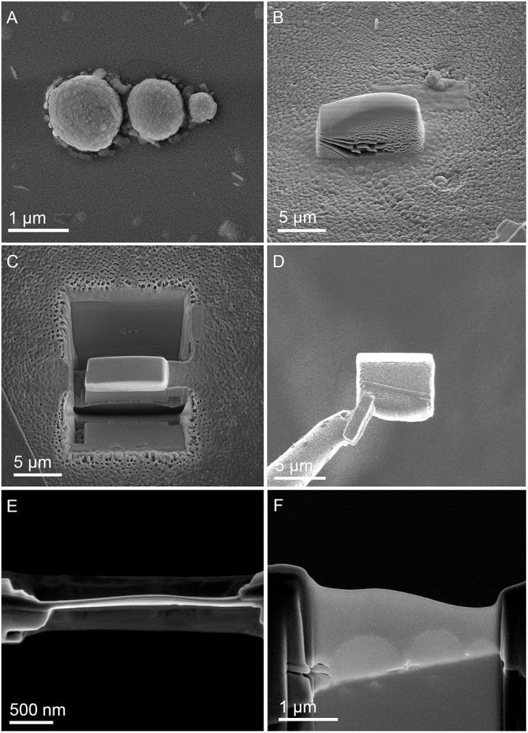
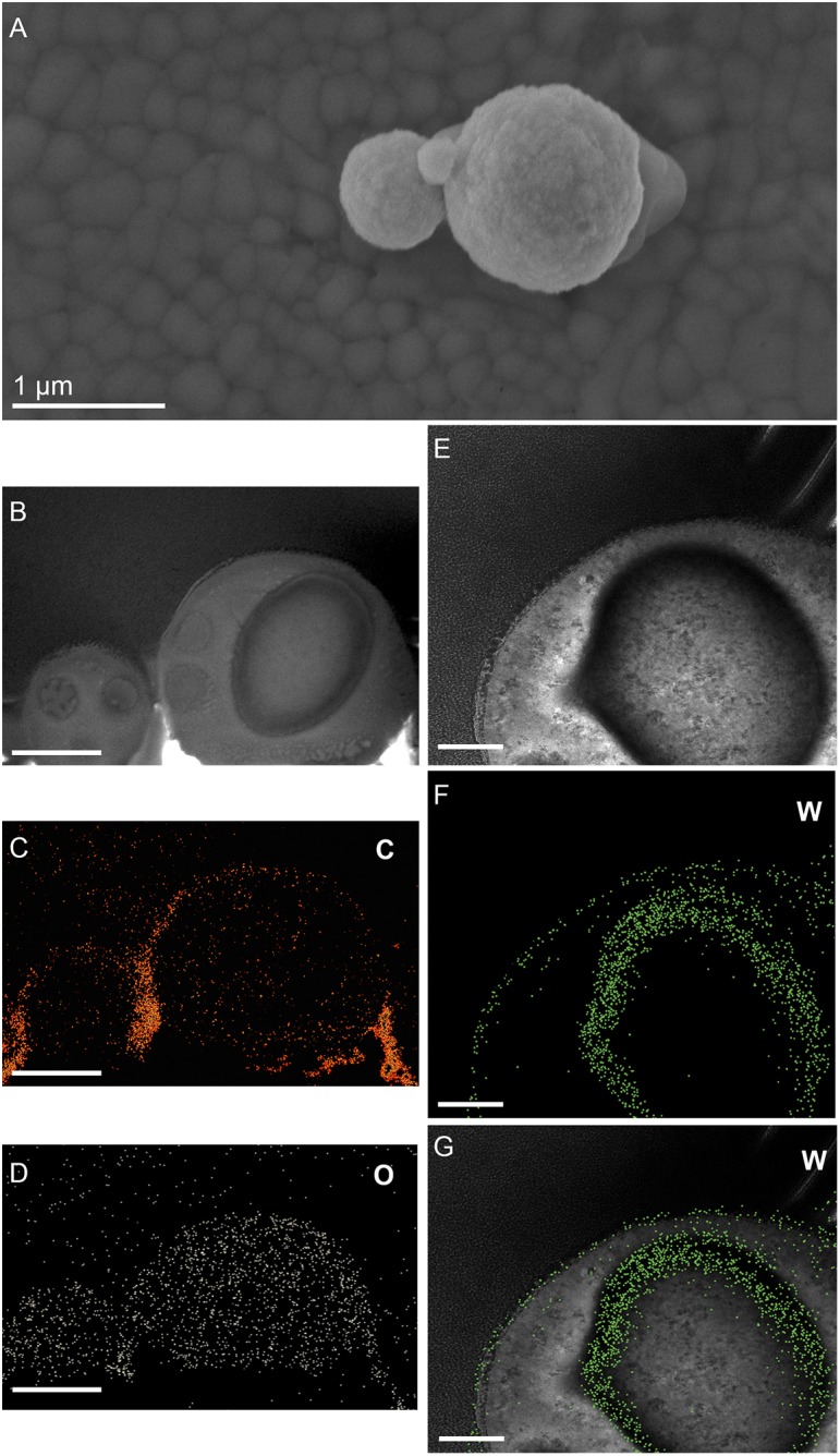
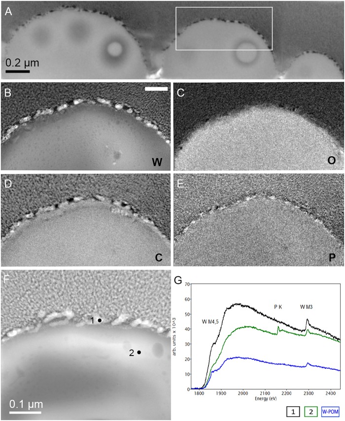
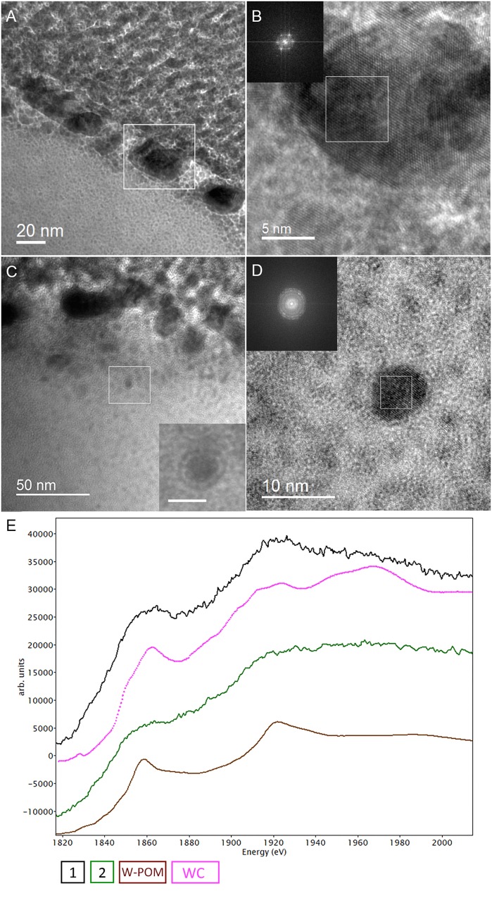
Inorganic systems based upon polyoxometalate (POM) clusters provide an experimental approach to develop artificial life. These artificial symmetric anionic macromolecules with oxidometalate polyhedra as building blocks were shown to be well suited as inorganic frameworks for complex self-assembling and organizing systems with emergent properties. Analogously to mineral cells based on iron sulfides, POMs are considered as inorganic cells in facilitating prelife chemical processes and displaying "life-like" characteristics. However, the relevance of POMs to life-sustaining processes (e.g., microbial respiration) has not yet been addressed, while iron sulfides are very well known as ubiquitous mineral precursors and energy sources for chemolithotrophic metabolism. Metallosphaera sedula is an extreme metallophilic and thermoacidophilic archaeon, which flourishes in hot acid and respires by metal oxidation. In the present study we provide our observations on M. sedula cultivated on tungsten polyoxometalate (W-POM). The decomposition of W-POM macromolecular clusters and the appearance of low molecular weight W species (e.g., WO) in the presence of M. sedula have been detected by electrospray ionization mass spectrometry (ESI-MS) analysis. Here, we document the presence of metalloorganic assemblages at the interface between M. sedula and W-POM resolved down to the nanometer scale using scanning and transmission electron microscopy (SEM and TEM) coupled to electron energy loss spectroscopy (EELS). High-resolution TEM (HR-TEM) and selected-area electron diffraction (SAED) patterns indicated the deposition of redox heterogeneous tungsten species on the S-layer of M. sedula along with the accumulation of intracellular tungsten-bearing nanoparticles, i.e., clusters of tungsten atoms. These results reveal the effectiveness of the analytical spectroscopy coupled to the wet chemistry approach as a tool in the analysis of metal-microbial interactions and microbial cultivation on supramolecular self-assemblages based on inorganic metal clusters. We discuss the possible mechanism of W-POM decomposition by M. sedula in light of unique electrochemical properties of POMs. The findings presented herein highlight unique metallophilicity in hostile environments, extending our knowledge of the relevance of POMs to life-sustaining processes, understanding of the transition of POMs as inorganic prebiotic model to life-sustainable material precursors and revealing biogenic signatures obtained after the decomposition of an artificial inorganic compound, which previously was not associated with any living matter.

Keywords: Metallosphaera sedula; archaea; biomineralisation; microbe–mineral interactions; tungsten.


Figures

FIGURE 1
FIGURE 1
Scanning electron microscopy (SEM) images showing cells of M. sedula after 21 days of cultivation with W-POM. (A) SEM image of colonies of M. sedula cells showing single cells of M. sedula connected by means of extracellular extensions. (B) Magnified SEM of the cellular assemblage, revealing the network of filamentous membrane extensions (the diameter in a size range of 10–30 nm) between the neighborhood cells of M. sedula. A number of nano-sized vesicles are localized in the areas surrounding the single cells and along the extracellular extensions.
FIGURE 2
FIGURE 2
Scanning electron microscope images of M. sedula cells and ESI-MS patters after 21 days of cultivation with W-POM. (A–E) SEM images showing various assemblages of dividing M. sedula cells and cells forming budding vesicles. (F) Experimental ESI-MS analysis of biogenic cultures of M. sedula (red pattern) and corresponding abiotic control comprised of non-inoculated growth medium (black patter) at the “0” time point and after 21 days of cultivation with W-POM.
FIGURE 3
FIGURE 3
Negative ion-mode ESI-MS spectrum after 0 and 21 days of cultivation with W-POM in mixed H2O/CH3CN (A) and corresponding species assigned to the peaks in the ESI-MS spectrum in panel (A,B).
FIGURE 4
FIGURE 4
The region of 860–890 m/z from the negative ion-mode ESI-MS spectra after 0 and 21 days of M. sedula cultivation with W-POM (Figure 3).
FIGURE 5
FIGURE 5
Focused Ion Beam (FIB) assisted preparation of thin lamellae of M. sedula cultivated on W-POM documented by electron beam induced SEM images. (A) SEM image of a cell assemblage of M. sedula used for milling. (B) 3 μm thick Pt deposition layer covering cells of M. sedula. (C) FIB removal of material at both sides of the Pt layer viewed perpendicular to the substrate surface. (D) Transfer of the 2.5 μm thick lamella from the micromanipulator needle (left) to the Cu TEM grid. (E) Finally thinned lamella showing the Pt layer in top-view. (F) Side-view of the finally thinned lamella showing the three flattened cells covered by a Pt layer.
FIGURE 6
FIGURE 6
Elemental ultrastructural analysis of cells of M. sedula after 21 days of cultivation with W-POM. (A) Transmission electron microscopy image showing an assemblage of the cells of M. sedula with the selected area used for energy-filtered transmission electron microscopy (EFTEM) analysis. (B–E) Corresponding tungsten (W), oxygen (O), carbon (C), and phosphorous (P) elemental maps. (F) The high angular annular dark field (HAADF) scanning TEM (STEM) image of a cell fragment of M. sedula with the depicted areas for W M3,4,5–edge core electron energy loss spectra (EELS) analysis. (G) Corresponding representative W M3,4,5–edge core EELS of W-POM and EELS acquired from the areas depicted in panel (F).
FIGURE 7
FIGURE 7
Additional elemental ultrastructural analysis of cells of M. sedula after 21 days of cultivation with W-POM. (A) Scanning electron microscopy (SEM) image showing an assemblage of the cells of M. sedula used for transmission electron microscopy (TEM) analysis. (B) TEM image showing an assemblage of the cells of M. sedula (C,D) Corresponding carbon (C) and oxygen (O) elemental maps. (E) Magnified TEM image of a cell of M. sedula. (F,G) Corresponding tungsten (W) elemental map and overlaid image. Scale bars are 1 μm.
FIGURE 8
FIGURE 8
Analytical spectroscopy of biogenic tungsten deposits in the cells of M. sedula after 21 days of cultivation with W-POM. (A) High-resolution TEM (HR-TEM) image of S-layer fragment of M. sedula cell. (B) HR-TEM and Fast Fourier Transform (FFT) acquired from the tungsten crystalline cell surface deposits labeled in panel (A). (C) HR-TEM image of the intracellular part of M. sedula cell; inlet represents magnified labeled area showing that intracellular nanoparticle is surrounded by electron dense layer which may suggest the presence of membrane; (D) HR-TEM and FFT obtained from intracellular nanoparticle labeled in panel (C). Insets in panel (B,D) represent diffraction patterns consistent with the tungsten carbide WC and W2C phases. (E) Comparison of Energy Los Near Edge Spectra (ELNES) recorded from W-M4,5 edge with the reference spectra from a WC phase (pink line) and simulations of W-containing phases of W-POM (brown line) and of the areas depicted in Figure 5F (black and green lines).

References

    1. Albu M., Pal A., Gspan C., Picu R. C., Hofer F., Kothleitner G. (2016). Self-organized Sr leads to solid state twinning in nano-scaled eutectic Si phase. Sci. Rep. 6:31635. 10.1038/srep31635 - DOI - PMC - PubMed
    1. Andreesen J. R., Makdessi K. (2008). Tungsten, the surprisingly positively acting heavy metal element for prokaryotes. Ann. N.Y. Acad. Sci. 1125 215–229. 10.1196/annals.1419.003 - DOI - PubMed
    1. Ankudinov A., Ravel B. (1998). Real-space multiple-scattering calculation and interpretation of x-ray-absorption near-edge structure. Phys. Rev. B Condens. Matter Mater. Phys. 58 7565–7576. 10.1103/PhysRevB.58.7565 - DOI
    1. Auernik K. S., Kelly R. M. (2008). Identification of components of electron transport chains in the extremely thermoacidophilic crenarchaeon Metallosphaera sedula through iron and sulfur compound oxidation transcriptomes. Appl. Environ. Microbiol. 74 7723–7732. 10.1128/AEM.01545-08 - DOI - PMC - PubMed
    1. Auernik K. S., Kelly R. M. (2010). Impact of molecular hydrogen on chalcopyrite bioleaching by the extremely thermoacidophilic archaeon Metallosphaera sedula. Appl. Environ. Microbiol. 76 2668–2672. 10.1128/AEM.02016-09 - DOI - PMC - PubMed

LinkOut - more resources