. 2019 Jun 13:10:1367.
doi: 10.3389/fimmu.2019.01367. eCollection 2019.

MicroRNA-155 Controls T Helper Cell Activation During Viral Infection

Affiliations

MicroRNA-155 Controls T Helper Cell Activation During Viral Infection

Eliana Goncalves-Alves et al. Front Immunol. .

Abstract

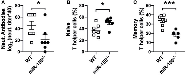
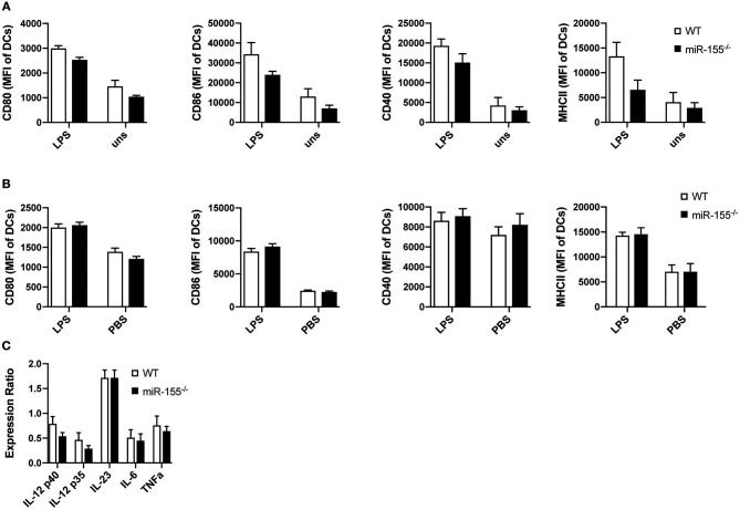
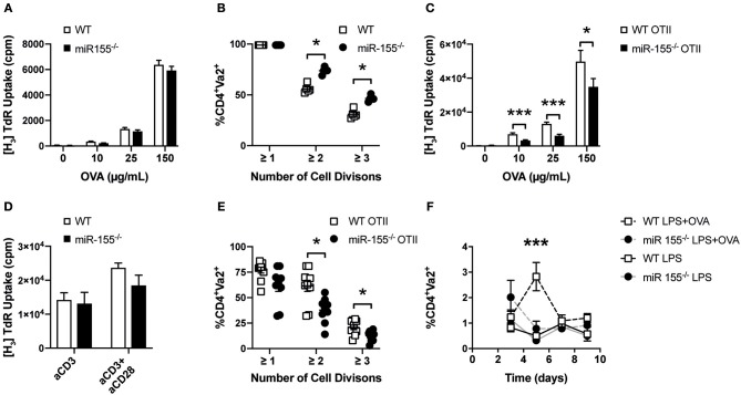
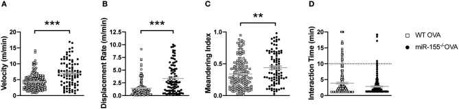
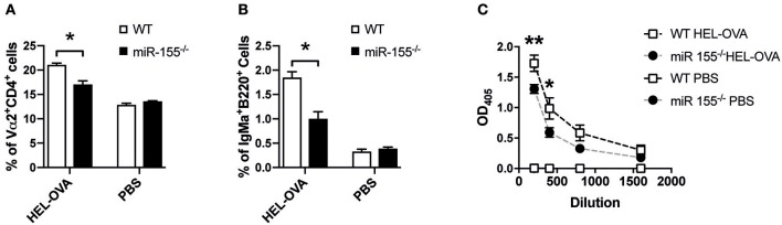
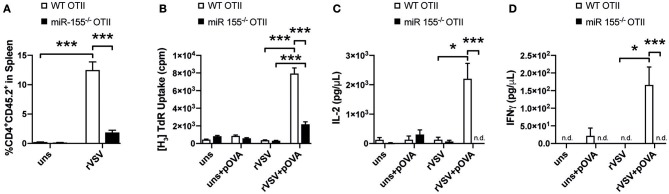
MicroRNA (miR) 155 has been implicated in the regulation of innate and adaptive immunity as well as autoimmune processes. Importantly, it has been shown to regulate several antiviral responses, but its contribution to the immune response against cytopathic viruses such as vesicular stomatitis virus (VSV) infections is not known. Using transgenic/recombinant VSV expressing ovalbumin, we show that miR-155 is crucially involved in regulating the T helper cell response against this virus. Our experiments indicate that miR-155 in CD4+ T cells controls their activation, proliferation, and cytokine production in vitro and in vivo upon immunization with OVA as well as during VSV viral infection. Using intravital multiphoton microscopy we analyzed the interaction of antigen presenting cells (APCs) and T cells after OVA immunization and found impaired complex formation when using miR-155 deficient CD4+ T cells compared to wildtype CD4+ T cells ex vivo. In contrast, miR-155 was dispensable for the maturation of myeloid APCs and for their T cell stimulatory capacity. Our data provide the first evidence that miR-155 is required for efficient CD4+ T cell activation during anti-viral defense by allowing robust APC-T cell interaction required for activation and cytokine production of virus specific T cells.

Keywords: APC-T cell interaction; APCs; T cell activation; T helper cells; antiviral immunity; microRNA-155.


Figures

Figure 1
Figure 1
Reduced host response to VSV infection in miR-155 deficient mice. (A) MiR-155−/− and WT mice were infected intravenously with Ovalbumin-expressing rVSV (2 × 10⁵ PFU/mL). After 8 days of infection, animals were sacrificed and serum and spleens were collected. Quantification of anti-VSV neutralizing antibodies present in mouse serum was done via VSV-specific plaque assay, as explained in material and methods Frequencies of naïve Th cells (CD62L+CD4+) (B) and memory Th cells (CD44+CD4+) (C) present in the spleen were quantified by flow cytometry. Data is presented as mean ± SEM, and representative of two independent experiments (n = 6–8 mice). Statistical significance was calculated using two-tailed Mann-Whitney test. *p ≤ 0.05, ***p < 0.001.
Figure 2
Figure 2
MiR-155 deficiency does not modify activation and function of DCs. (A) WT and miR-155−/− BMDCs cultured for 24 h in the presence or absence of LPS (10 μg/mL) were analyzed for the expression of CD80, CD86, CD40, and MHC II (MHC II) by flow cytometry. Bars show mean fluorescence intensity (MFI) ± SEM of CD11c+Gr-1 gated cells (n = 5 per condition, pooled from two independent experiments). (B) MFI ± SEM of the expression of CD80, CD86, CD40, MHC II of splenic CD11c+Gr-1 cells of WT and miR-155−/− mice 6 h after intraperitoneal injection of LPS (75 μg/mouse) or PBS (n = 5 per condition). (C) CD11c+Gr-1 cells were FACS sorted from spleens of mice treated with LPS for 6 h and mRNA expressions levels of the pro-inflammatory cytokines IL-12, IL-23, IL-6, and TNFα was quantified by qPCR (n = 5). Data is presented as mean ± SEM. Statistical significance was calculated using two-tailed Mann-Whitney test with Bonferroni correction.
Figure 3
Figure 3
Absence of miR-155 in CD4+ T cells leads to reduced clonal expansion. (A) WT or miR-155−/− BMDCs were co-cultured with WT OTII T cells in a 1:5 ratio in the presence of the indicated concentrations of OVA. Proliferation was quantified by H3-Thymidine incorporation. Results show mean values ± SEM of 6 technical replicates using two mice per genotype. The plot is representative of 3 independent experiments. (B) OTII T helper cells were stained with CFSE and transferred i.v. into WT and miR-155−/− mice. One day later mice were immunized i.v. with LPS (75 μg/mouse) and OVA (30 μg/mouse). Cell proliferation was quantified 3 days after immunization using flow cytometry by measuring CFSE mean fluorescence intensity of Vα2+ cells, gated on CD4+ cells in the spleen (n = 5 for each genotype). (C) WT or miR-155−/− OTII T cells were co-cultured with WT BMDCs in a 1:5 ratio in the presence of the indicated concentrations of OVA. Proliferation was quantified by H3-Thymidine incorporation. Results show mean values ± SEM of 6 technical replicates and two mice per genotype. The plot is representative of 3 independent experiments. (D) CD4+ cells were MACS isolated from spleen of WT or miR-155−/− mice and seeded in 96 well plates pre-coated with anti-CD3 (1 μg/mL) and anti-CD28 (3 μg/mL). Proliferation was quantified by H3-Thymidine incorporation. Results show mean values ± SEM of 6 technical replicates and two mice per genotype. The plot is representative of 3 independent experiments. (E) WT or miR-155−/− OTII T cells were stained with CFSE and transferred i.v. into WT. Mice were immunized i.v. with LPS (75 μg/mouse) with OVA (30 μg/mouse). Cell expansion was quantified 3 days after immunization using flow cytometry by measuring CFSE mean fluorescence intensity of Vα2+ cells, gated on CD4 + cells (n = 8 for each genotype). (F) WT or miR-155−/− OTII T helper cells were transferred i.v. into CD45.1 mice (immunization was done as referred above). Frequency of CD45.2+Vα2+ cells of gated CD4 + cells, present 3, 5, 7, or 9 days after immunization was done using flow cytometry (n = 3 per condition and genotype). Data is presented as mean ± SEM, statistical significance was calculated using two-tailed Mann-Whitney test with Bonferroni correction for all panels except (F) where Two-way ANOVA was used. Significance shown reflect comparison of WT vs. miR-155−/−. *p < 0.05, ***p < 0.001.
Figure 4
Figure 4
MiR-155 in CD4+ T cells is required for proper DC-T cell interactions. CD11c-YFP mice received 5 × 10⁶ WT or miR-155 OTII T cells labeled with CMTPX and were immediately immunized with OVA/CFA in the footpad. Popliteal LNs were collected 24 h after immunization, three random regions of each LN were imaged and Volocity software was used for semi-automated tracking. Each point represents a single tracked object, either a single cell or the interaction (intersection between the DC and T cell fluorescent signal). Mean velocity (A), displacement rate (B), meandering index (C), and interaction time between DCs and T cells (D) were quantified using Volocity software programs. Data are presented as mean ± SEM, two-tailed Mann-Whiney test and representative of two animals per group of at least two independent experiments. ***p ≤ 0.001, **p ≤ 0.01.
Figure 5
Figure 5
MiR-155 deficiency in T cells affects T cell help to B cells. (A) WT mice received 5 × 10⁶ HEL-specific B cells and 5 × 10⁶ WT or miR-155 OTII T cells and brachial LN were collected 6 days after immunization with HEL-OVA/CFA. Expansion of transferred OTII T cells was quantified by flow cytometry as frequency of Vα2+ cells gated on CD4+ cells. (B) Expansion of HEL-specific B cells was also analyzed by quantifying IgMa+ cells, gates on B220+ cells. (C) Serum was collected at experimental endpoint and assessed for the presence of anti-HEL IgMa by ELISA. Data are presented as mean ± SEM and representative of two different experiments (n = 6). Statistical significance was calculated using two-tailed Mann-Whitney test with Bonferroni correction in (A,B), while Two-way ANOVA was used in (C). *p ≤ 0.05, **p ≤ 0.01.
Figure 6
Figure 6
MiR-155 deficiency diminishes T helper cell activation during viral infection. (A) CD45.1 mice received 3 × 10⁶ WT or miR-155 or WT OTII T cells, mice were infected i.v. with rVSV the following day. Spleens were collected 8 days after infection. Expansion of transferred cells was quantified as percentage of CD45.2+ from all CD4+ cells. (B) Splenocytes were cultured for 3 days in full RPMI with or without OVA peptide (323–339) and proliferation was quantified by H3-Thymidine incorporation. Flow Cytomix was used to quantify levels of IL-2 (C) and IFN-γ (D) present in the supernatant of splenocyte re-stimulation cultures. Data is presented as mean ± SEM and representative of two independent experiments (n = 6). Statistical significance was calculated using two-tailed Mann-Whitney test with Bonferroni correction. *p ≤ 0.05; ***p ≤ 0.001.

Similar articles

Cited by

References

    1. Elton TS, Selemon H, Elton SM, Parinandi NL. Regulation of the MIR155 host gene in physiological and pathological processes. Gene. (2013) 532:1–12. 10.1016/j.gene.2012.12.009 - DOI - PubMed
    1. Vigorito E, Kohlhaas S, Lu D, Leyland R. miR-155: an ancient regulator of the immune system. Immunol Rev. (2013) 253:146–57. 10.1111/imr.12057 - DOI - PubMed
    1. Alivernini S, Gremese E, Mcsharry C, Tolusso B, Ferraccioli G, Mcinnes IB, et al. . MicroRNA-155-at the critical interface of innate and adaptive immunity in arthritis. Front Immunol. (2017) 8:1932. 10.3389/fimmu.2017.01932 - DOI - PMC - PubMed
    1. Ceppi M, Pereira PM, Dunand-Sauthier I, Barras E, Reith W, Santos MA, et al. . MicroRNA-155 modulates the interleukin-1 signaling pathway in activated human monocyte-derived dendritic cells. Proc Natl Acad Sci USA. (2009) 106:2735–40. 10.1073/pnas.0811073106 - DOI - PMC - PubMed
    1. Dunand-Sauthier I, Santiago-Raber ML, Capponi L, Vejnar CE, Schaad O, Irla M, et al. . Silencing of c-Fos expression by microRNA-155 is critical for dendritic cell maturation and function. Blood. (2011) 117:4490–500. 10.1182/blood-2010-09-308064 - DOI - PubMed

Publication types

MeSH terms