Review
. 2019 Jun 2:2019:3706315.
doi: 10.1155/2019/3706315. eCollection 2019.

Skin Acute Wound Healing: A Comprehensive Review

Affiliations
Review

Skin Acute Wound Healing: A Comprehensive Review

Luis Cañedo-Dorantes et al. Int J Inflam. .

Abstract

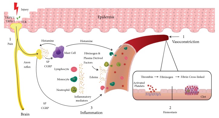
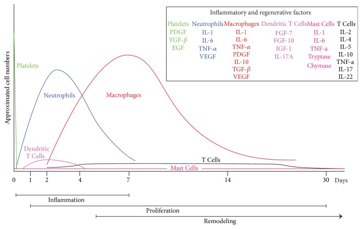
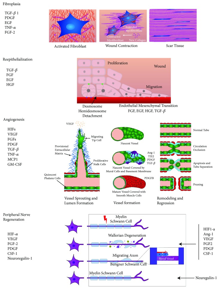
Experimental work of the last two decades has revealed the general steps of the wound healing process. This complex network has been organized in three sequential and overlapping steps. The first step of the inflammatory phase is an immediate response to injury; primary sensory neurons sense injury and send danger signals to the brain, to stop bleeding and start inflammation. The following target of the inflammatory phase, led by the peripheral blood mononuclear cells, is to eliminate the pathogens and clean the wound. Once this is completed, the inflammatory phase is resolved and homeostasis is restored. The aim of the proliferative phase, the second phase, is to repair wound damage and begin tissue remodeling. Fibroplasia, reepithelialization, angiogenesis, and peripheral nerve repair are the central actions of this phase. Lastly, the objective of the final phase is to complete tissue remodeling and restore skin integrity. This review provides present day information regarding the status of the participant cells, extracellular matrix, cytokines, chemokines, and growth factors, as well as their interactions with the microenvironment during the wound healing process.


Figures

Figure 1
Figure 1
See text.
Figure 2
Figure 2
Neutrophil accumulation in the wound increases during the initial inflammatory phase and starts declining 4 days later until the end of the week [49]. Macrophages increase their numbers during the inflammation phase, reach maximum concentration during the proliferation phase, and decline progressively during the remodeling phase, being the most abundant cell in all phases of wound repair [63, 115]. Lymphocytes start increasing their number after injury and reach a plateau at day 4 that continues to be present until the last phase. The skin resident cell number in dendritic cells, mast cells, Trm, and Treg rm is not known, but the presence in the skin of a large population of T memory cells protects the skin against reinfection [99]; further research will be required to clarify their functions in the wound healing process. The approximated time of each of the wound healing phases is illustrated in the bars at the bottom of the graphic [41].
Figure 3
Figure 3
See Sections 5.4.1to 5.4.4.

References

    1. Nejati R., Kovacic D., Slominski A. Neuro-Immune-Endocrine Functions of The Skin: An Overview. Taylor & Francis; 2013. - PMC - PubMed
    1. Pasparakis M., Haase I., Nestle F. O. Mechanisms regulating skin immunity and inflammation. Nature Reviews Immunology. 2014;14(5):289–301. doi: 10.1038/nri3646. - DOI - PubMed
    1. Rousselle P., Braye F., Dayan G. Re-epithelialization of adult skin wounds: cellular mechanisms and therapeutic strategies. Advanced Drug Delivery Reviews. 2018 - PubMed
    1. Kupper T. S., Fuhlbrigge R. C. Immune surveillance in the skin: mechanisms and clinical consequences. Nature Reviews Immunology. 2004;4(3):211–222. doi: 10.1038/nri1310. - DOI - PMC - PubMed
    1. Ebenezer G., Polydefkis M. Epidermal innervation in diabetes. Handbook of Clinical Neurology. 2014;126:261–274. doi: 10.1016/B978-0-444-53480-4.00020-5. - DOI - PubMed

LinkOut - more resources