Review
. 2019 Jun;9(3):281-298.
doi: 10.21037/cdt.2019.04.03.

Current state-of-play in spontaneous coronary artery dissection

Affiliations
Review

Current state-of-play in spontaneous coronary artery dissection

Kyle B Franke et al. Cardiovasc Diagn Ther. 2019 Jun.

Abstract

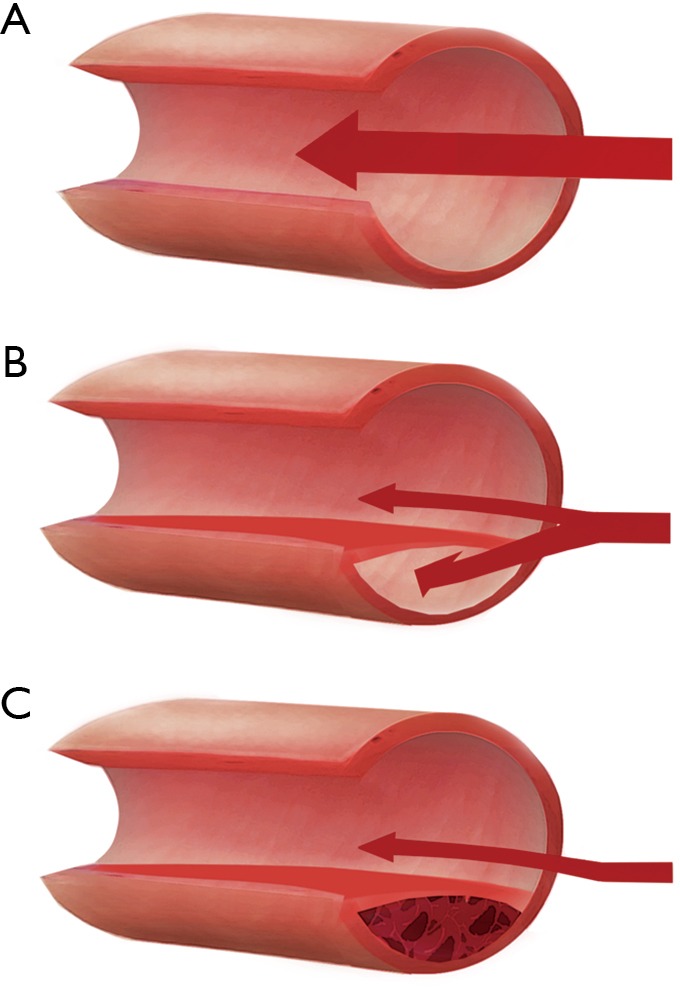
For over 80 years, spontaneous coronary artery dissection (SCAD) has been recognised as a cause of myocardial infarction. SCAD is described as a non-iatrogenic, non-atherosclerotic coronary artery dissection, resulting in formation of a false lumen or intramural haematoma in the coronary artery wall that compresses the true lumen, often compromising myocardial blood flow. In early literature, the incidence of SCAD in acute coronary syndrome (ACS) was underestimated. Recent advances in awareness and widespread early angiographic investigation in ACS has led to important shifts in our understanding of the prevalence, predisposing causes, natural history, aetiology, clinical and angiographic features, management, and prognosis of SCAD. It is now well understood that SCAD predominantly affects women and is responsible for around 20% of ACS presentations in females below the age of 60. Despite this, SCAD is still often overlooked and misdiagnosed as atherosclerotic disease. Misdiagnosis is multifactorial; with contributing factors including a low clinical index of suspicion, particularly in young females, a lack of clinician familiarity with angiographic variants, and limitations of angiography. Although increasing evidence suggests that optimal management is distinct from atherosclerotic coronary artery disease, many questions remain unanswered regarding the pathogenesis and optimal treatment of SCAD, heralding prospective research to answer these questions. This review aims to give a current clinical perspective on SCAD and highlight the importance of familiarity and vigilance with this condition when diagnosing and treating ACS.

Keywords: Acute coronary syndrome (ACS); myocardial infarction; non-atherosclerotic coronary artery disease; spontaneous coronary artery dissection (SCAD).


Conflict of interest statement

Conflicts of Interest: The authors have no conflicts of interest to declare.
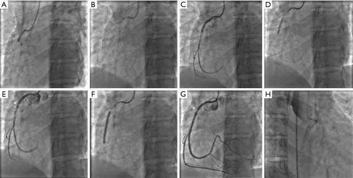
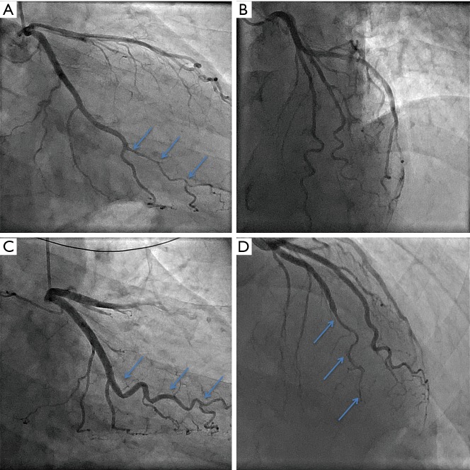
Figures

Figure 1
Figure 1
Pathological mechanisms underlying SCAD. Illustrations are shown of (A) a normal coronary artery, (B) intimal tear, resulting in blood flow under the tunica intima with creation of a false lumen restricting blood flow, and (C) an intramural haematoma which compresses the true lumen of the artery. In cases of intimal tear, it is still unclear if this is typically the primary event, or secondary to intramural haematoma. SCAD, spontaneous coronary artery dissection.
Figure 2
Figure 2
Angiographic classification of SCAD. (A) shows Type 1 SCAD of the right coronary artery, characterised by a double lumen illustrated by contrast hold-up. (B) and (C) show Type 2 SCAD of the left anterior descending artery, which involves abrupt narrowing of the coronary artery with a diffuse tubular stenosis, either for a section of the artery in Type 2a (B), or to the distal end of the artery in Type 2b (C). (D) shows Type 3 SCAD of the second obtuse marginal branch of the left circumflex artery, mimicking atherosclerotic disease. In this case, SCAD was confirmed by optical coherence tomography. Asterisks denote the locations of dissection. SCAD, spontaneous coronary artery dissection.
Figure 3
Figure 3
Optical coherence tomography confirmation of SCAD. (A) Angiographic appearance of Type 3 SCAD of the left anterior descending artery (arrows), confirmed with (B) optical coherence tomography imaging, showing intramural haematoma (H) and false lumen (FL). (C) Angiogram image of right coronary artery in patient who presented with acute coronary syndrome showing beaded appearance (arrow) and (D) accompanying optical coherence tomography image showing an intimal flap (arrow) and false lumen (FL). SCAD, spontaneous coronary artery dissection.
Figure 4
Figure 4
Spontaneous healing of SCAD. (A) shows Type 2b SCAD detected on coronary angiogram of the obtuse marginal branch of a left circumflex artery (arrows), which was treated conservatively. (B) shows complete healing of the same artery (arrows) three months later on a follow-up angiogram that was performed for recurrent chest pain without biomarker elevation. SCAD, spontaneous coronary artery dissection.
Figure 5
Figure 5
Complication from PCI of possible unrecognised SCAD. A 42-year-old woman presented with an inferior STEMI in the absence of known atherosclerotic risk factors. (A) Emergency coronary angiography showed an occlusion of the right coronary artery. (B) The treating interventionalist used an aggressive guide catheter (6F AL1) and successfully wired the occlusion. (C) This restored TIMI 2/3 flow and revealed a tubular stenosis. The possibility of a Type 2b SCAD was not recognised and the case proceeded to angioplasty and stenting. (D) Balloon pre-dilation was performed, leaving in (E) what appears to be a small dissection proximal to the previous stenosis (*). (F) A long stent was deployed, but as shown in (G) did not cover the proximal dissection (*). Although this was noted, a decision was made to not cover this lesion and the guide wire was pulled back into the proximal artery. (H) shows complete closure of the vessel immediately after, with dissection now extending back to the origin of the right coronary artery and into the aortic root and ascending aorta. Further attempts at salvage PCI were unsuccessful and emergency surgery was required. PCI, percutaneous coronary intervention; SCAD, spontaneous coronary artery dissection.
Figure 6
Figure 6
Recurrent SCAD. (A) and (B) are angiographic images taken of a 52-year-old man who initially presented with a NSTEMI in the absence of traditional atherosclerosis risk factors. At the time, his angiogram was reported as showing minor luminal irregularities of all epicardial coronary arteries, and the treating team remained uncertain as to the cause of his presentation. He re-presented with a second NSTEMI six months later, with repeat angiographic images shown in (C) and (D). On this occasion, SCAD was identified in the mid-distal left anterior descending artery, denoted by arrows in (D). In retrospect, it was also recognised that there had been SCAD in the obtuse marginal branch of the circumflex artery at the time of the first presentation, denoted by arrows in (A), and this had now healed as indicated by arrows in (C). Note also, the tortuosity of both SCAD-affected vessels. NSTEMI, non-ST segment elevation myocardial infarction; SCAD, spontaneous coronary artery dissection.

References

    1. Pretty HC. Dissecting aneurysm of coronary artery in a woman aged 42: rupture. Br Med J 1931;1:667.
    1. Kamineni R, Sadhu A, Alpert JS. Spontaneous coronary artery dissection: report of two cases and a 50-year review of the literature. Cardiol Rev 2002;10:279-84. 10.1097/00045415-200209000-00004 - DOI - PubMed
    1. Saw J, Mancini GBJ, Humphries KH. Contemporary Review on Spontaneous Coronary Artery Dissection. J Am Coll Cardiol 2016;68:297-312. 10.1016/j.jacc.2016.05.034 - DOI - PubMed
    1. Adlam D, Alfonso F, Maas A, et al. European Society of Cardiology, acute cardiovascular care association, SCAD study group: a position paper on spontaneous coronary artery dissection. Eur Heart J 2018;39:3353-68. 10.1093/eurheartj/ehy080 - DOI - PMC - PubMed
    1. Hayes SN, Kim ESH, Saw J, et al. Spontaneous Coronary Artery Dissection: Current State of the Science: A Scientific Statement From the American Heart Association. Circulation 2018;137:e523-57. 10.1161/CIR.0000000000000564 - DOI - PMC - PubMed