. 2019 Jul 5;14(7):e0219280.
doi: 10.1371/journal.pone.0219280. eCollection 2019.

Loss of function mutation of Eftud2, the gene responsible for mandibulofacial dysostosis with microcephaly (MFDM), leads to pre-implantation arrest in mouse

Affiliations

Loss of function mutation of Eftud2, the gene responsible for mandibulofacial dysostosis with microcephaly (MFDM), leads to pre-implantation arrest in mouse

Marie-Claude Beauchamp et al. PLoS One. .

Abstract

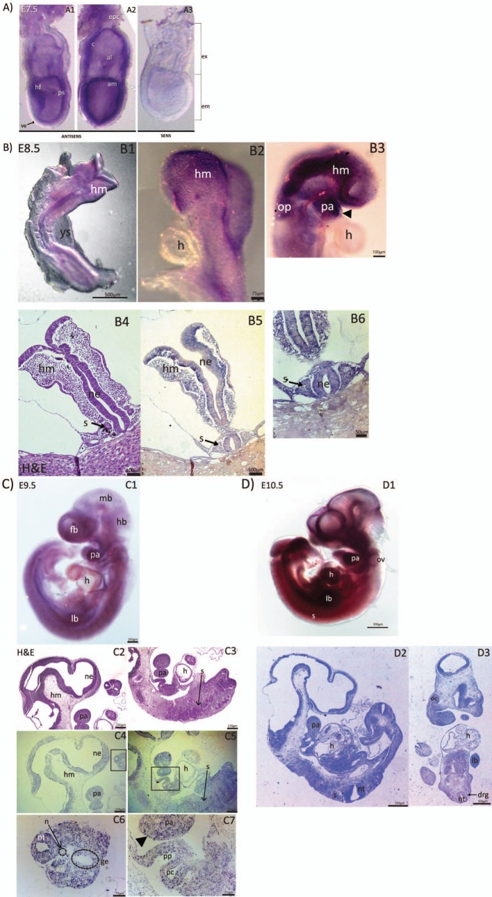
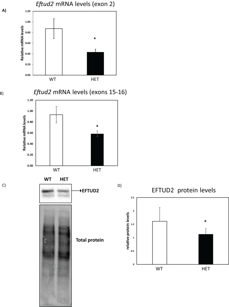
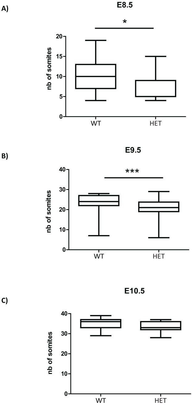
Mutations in EFTUD2 are responsible for the autosomal dominant syndrome named MFDM (mandibulofacial dysostosis with microcephaly). However, it is not clear how reduced levels of EFTUD2 cause abnormalities associated with this syndrome. To determine if the mouse can serve as a model for uncovering the etiology of abnormalities found in MFDM patients, we used in situ hybridization to characterize expression of Eftud2 during mouse development, and used CRISPR/Cas9 to generate a mutant mouse line with deletion of exon 2 of the mouse gene. We found that Eftud2 was expressed throughout embryonic development, though its expression was enriched in the developing head and craniofacial regions. Additionally, Eftud2 heterozygous mutant embryos had reduced EFTUD2 mRNA and protein levels. Moreover, Eftud2 heterozygous embryos were born at the expected Mendelian frequency, and were viable and fertile despite being developmentally delayed. In contrast, Eftud2 homozygous mutant embryos were not found post-implantation but were present at the expected Mendelian frequency at embryonic day (E) 3.5. Furthermore, only wild-type and heterozygous E3.5 embryos survived ex vivo culture. Our data indicate that Eftud2 expression is enriched in the precusor of structures affected in MFDM patients and show that heterozygous mice carrying deletion of exon 2 do not model MFDM. In addition, we uncovered a requirement for normal levels of Eftud2 for survival of pre-implantation zygotes.


Conflict of interest statement

The authors have declared that no competing interests exist.

Figures

Fig 1
Fig 1. Representative images from in situ hybridation (ISH) showing Eftud2 expression in wild-type CD1 embryos at various developmental stages.
A) Wholemount ISH on E7.5 embryos using antisense (A1-A2) and sense probes (A3). Eftud2 is expressed in both embryonic (em) and extra-embryonic (ex) region. Eftud2 is found in headfold (hf), primitive streak (ps), amnion (am), allantois (al), chorion (c) and ectoplacental cone (epc), but not in visceral endoderm (ve). B) Wholemount and section ISH on E8.5 embryos. Eftud2 was expressed in head mesenchyme (hm), pharyngeal arches (pa) and otic placode (op) (B1-B3). No expression was found in the heart (h) (B2-B3). Headarrow points to the ectoderm of pa (B3). H&E staining of region examined in B5 (B4). Sections ISH reveals expression in head mesenchyme (hm), somites (s) and neural epithelium (ne) (B5-B6). C) Wholemount (C1) and ISH on sagittal sections (C4-C7) of E9.5 embryos. Expression was found in the forebrain (fb), midbrain (mb), hindbrain (hb), pharyngeal arches (pa) and limb buds (lb) while reduced expression was found in the heart (h) (C1). H&E staining of regions stained is shown (C2-C3). ISH on sagittal sections shows enriched expression in neural epithelium (ne) and pharyngeal arches (pa) (C4). Reduced expression was found in the heart (h), and the somite (s) (C5). Higher magnification image of the boxed region in C4 indicate that Eftud2 was also expressed in the neural tube (nt) and gut epithelium (ge) but not in the notochord (n) (C6). Higher magnification image of the boxed region in C5 shows expression in pharyngeal pouches (pp) and paryngeal clefts (pc) and in non-neural ectoderm (arrowhead) (C7). D) Wholemount (D1), ISH on sagittal (D2) and frontal (D3) sections of E10.5 embryos. At this stage, expression was ubiquitous and included otic vesicles (ov), dorsal root ganglia (drg) and optic cup inner layer (oc).
Fig 2
Fig 2. Reduced Eftud2 mRNA and protein levels in heterozygous mice.
Eftud2 mRNA levels was evaluated using RT-qPCR in E9.5 embryos on the mixed genetic background with primers flanking A) exon 2 or B) exons 15–16. WT = 3, HET = 3 (see Mat&Methods section for samples description). C) Total proteins from E11.5 embryos on the mixed genetic background were subjected to Western blot as described in the Mat&Methods section. One representative blot of 3 independent experiments is shown and D) quantification of EFTUD2 relative to total protein level. WT = 5, HET = 15. Results represent average ± SD. *P<0.05 by t-test.
Fig 3
Fig 3. Eftud2 heterozygous embryos on the mixed CD1:FVB genetic background have reduced number of somites at E8.5 and E9.5.
Somites counted at A) E8.5 WT = 11; HET = 27 B) E9.5, WT = 32; HET = 72 C) E10.5, WT = 15; HET = 40. *P<0.05, ***P<0.001 by t-test.

References

    1. Lehalle D, Gordon CT, Oufadem M, Goudefroye G, Boutaud L, Alessandri JL, et al. Delineation of EFTUD2 haploinsufficiency-related phenotypes through a series of 36 patients. Human mutation. 2014;35(4):478–85. Epub 2014/01/29. 10.1002/humu.22517 . - DOI - PubMed
    1. Sarkar A, Emrick LT, Smith EM, Austin EG, Yang Y, Hunter JV, et al. Novel de novo mutations in EFTUD2 detected by exome sequencing in mandibulofacial dysostosis with Microcephaly syndrome. American journal of medical genetics Part A. 2015;167a(4):914–8. Epub 2015/03/05. 10.1002/ajmg.a.36948 . - DOI - PubMed
    1. Rengasamy Venugopalan S, Farrow EG, Lypka M. Whole-exome sequencing identified a variant in EFTUD2 gene in establishing a genetic diagnosis. Orthodontics & craniofacial research. 2017;20 Suppl 1:50–6. Epub 2017/06/24. 10.1111/ocr.12150 . - DOI - PubMed
    1. Huang L, Vanstone MR, Hartley T, Osmond M, Barrowman N, Allanson J, et al. Mandibulofacial Dysostosis with Microcephaly: Mutation and Database Update. Human mutation. 2016;37(2):148–54. Epub 2015/10/29. 10.1002/humu.22924 - DOI - PMC - PubMed
    1. Yu KPT, Luk HM, Gordon CT, Fung G, Oufadem M, Garcia-Barcelo MM, et al. Mandibulofacial dysostosis Guion-Almeida type caused by novel EFTUD2 splice site variants in two Asian children. Clinical dysmorphology. 2018;27(2):31–5. Epub 2018/01/31. 10.1097/MCD.0000000000000214 . - DOI - PubMed

Publication types

Substances

Grants and funding

LinkOut - more resources