Review
. 2019 Sep;28(9):1567-1581.
doi: 10.1002/pro.3674. Epub 2019 Aug 6.

β-Amyloid aggregation and heterogeneous nucleation

Affiliations
Review

β-Amyloid aggregation and heterogeneous nucleation

Atul K Srivastava et al. Protein Sci. 2019 Sep.

Abstract

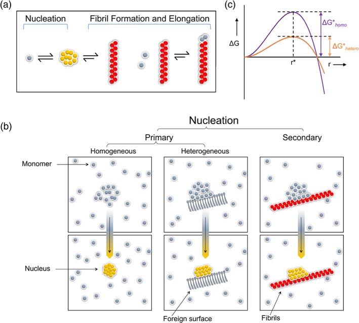
In this article, we consider the role of heterogeneous nucleation in β-amyloid aggregation. Heterogeneous nucleation is more common and occurs at lower levels of supersaturation than homogeneous nucleation. The nucleation period is also the stage at which most of the polymorphism of amyloids arises, this being one of the defining features of amyloids. We focus on several well-known heterogeneous nucleators of β-amyloid, including lipid surfaces, especially those enriched in gangliosides and cholesterol, and divalent metal ions. These two broad classes of nucleators affect β-amyloid particularly in light of the amphiphilicity of these peptides: the N-terminal region, which is largely polar and charged, contains the metal binding site, whereas the C-terminal region is aliphatic and is important in lipid binding. Notably, these two classes of nucleators can interact cooperatively, aggregation begetting greater aggregation.

Keywords: Alzheimer disease; amyloid-β (Aβ) aggregation; cholesterol; gangliosides; heterogeneous nucleation; lipids; metal ions.


Figures

Figure 1
Figure 1
Homogeneous and heterogeneous nucleation. (a) In nucleation (for crystallization or fibrillization), the rate limiting step is the formation of a multimolecular nucleus, defined as the smallest aggregate for which the free energy for adding subunits (leading to formation of the crystal or fibril) is lower than that of losing subunits (leading to dissolution). This process is thermodynamically distinct from fibril growth (or elongation). (b) In homogeneous nucleation, the nucleus is composed of only aggregating molecules. Heterogeneous nucleation occurs on a foreign surface. (c) As described in the text, heterogeneous nucleation is generally energetically more favorable than homogeneous nucleation and thus is more common
Figure 2
Figure 2
The amphiphilicity of Aβ peptides. (a) Sequence of human Aβ (Aβ42 is shown), color‐coded. (Hydrophobic residues are yellow; charged/polar residues are red, blue, green for acidic, basic, uncharged, respectively. Hydrophobicity scales differ: Tyr is considered here as hydrophobic; His is considered as polar and uncharged, though obviously it bears partial positive charge at near neutral pH; and Gly residues are depicted in black). The hydrophobic (or lipophilic) residues form two clusters, residues 17–21 and 30–42, which eventually form most of the two β‐sheet segments in fibrils. (b) Formation of Aβ aggregates on “ganglioside nanoclusters” was from mouse neuronal lipids, including mainly gangliosides, sphingomyelin (SM), and cholesterol. Morphology of these aggregates depended strongly on lipid composition. Reprinted from Reference 35
Figure 3
Figure 3
Three structures of Aβ peptides binding metal ions. (a) Human‐Aβ1‐16 monomer (Zn2+); pdb 1ze9. This peptide, lacking the hydrophobic domains, does not form fibrils and can exist at higher concentrations in solution than full length Aβ peptides. Here, this peptide is shown as a monomer binding Zn2+. (b) Human‐Aβ(1‐16) dimer (Zn2+); pdb 2mgt. This same peptide, at different Aβ:Zn2+ ratios, forms a dimer bridged by a Zn2+ ion, the “English mutation” of Aβ (H6R).155 (c) Rat‐Aβ(1‐16) dimer (Zn2+); pdb 2li9. The rat Aβ(1‐16) peptide also binds Zn2+ and other metal ions, despite lacking the His13 residue of human Aβ
Figure 4
Figure 4
Because of the amphiphilicity of Aβ peptides, different modes of heterogeneous nucleation can work together and amplify one another. Metal ions lead to formation of Aβ dimers or oligomers (a), which also brings the hydrophobic C‐termini into proximity and concentrates them (b), leading to further aggregation. (c) Aβ peptides, whether metal‐bound or not, have hydrophobic C‐termini that can bind to lipid surfaces

References

    1. Hellstrand E, Nowacka A, Topgaard D, Linse S, Sparr E. Membrane lipid co‐aggregation with alpha‐synuclein fibrils. PLoS One. 2013;8:e77235. - PMC - PubMed
    1. Iyer A, Petersen NO, Claessens MM, Subramaniam V. Amyloids of alpha‐synuclein affect the structure and dynamics of supported lipid bilayers. Biophys J. 2014;106:2585–2594. - PMC - PubMed
    1. Last NB, Rhoades E, Miranker AD. Islet amyloid polypeptide demonstrates a persistent capacity to disrupt membrane integrity. Proc Natl Acad Sci U S A. 2011;108:9460–9465. - PMC - PubMed
    1. Schlamadinger DE, Miranker AD. Fiber‐dependent and ‐independent toxicity of islet amyloid polypeptide. Biophys J. 2014;107:2559–2566. - PMC - PubMed
    1. Bender ML, Kezdy J. Mechanism of action of proteolytic enzymes. Annu Rev Biochem. 1965;34:49–76. - PubMed

Publication types

LinkOut - more resources