. 2019 Sep:26:101237.
doi: 10.1016/j.redox.2019.101237. Epub 2019 Jun 5.

Extracellular vesicle-mediated macrophage activation: An insight into the mechanism of thioredoxin-mediated immune activation

Affiliations

Extracellular vesicle-mediated macrophage activation: An insight into the mechanism of thioredoxin-mediated immune activation

Chontida Yarana et al. Redox Biol. 2019 Sep.

Abstract

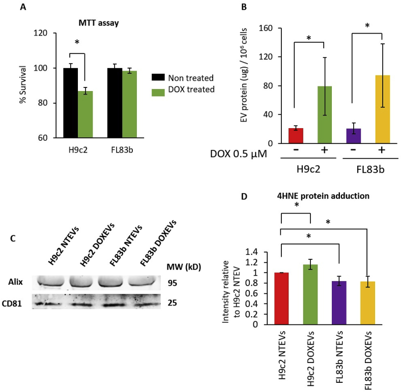
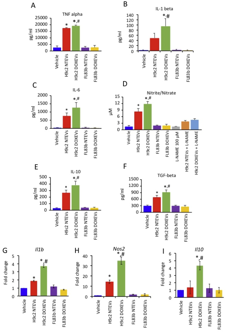
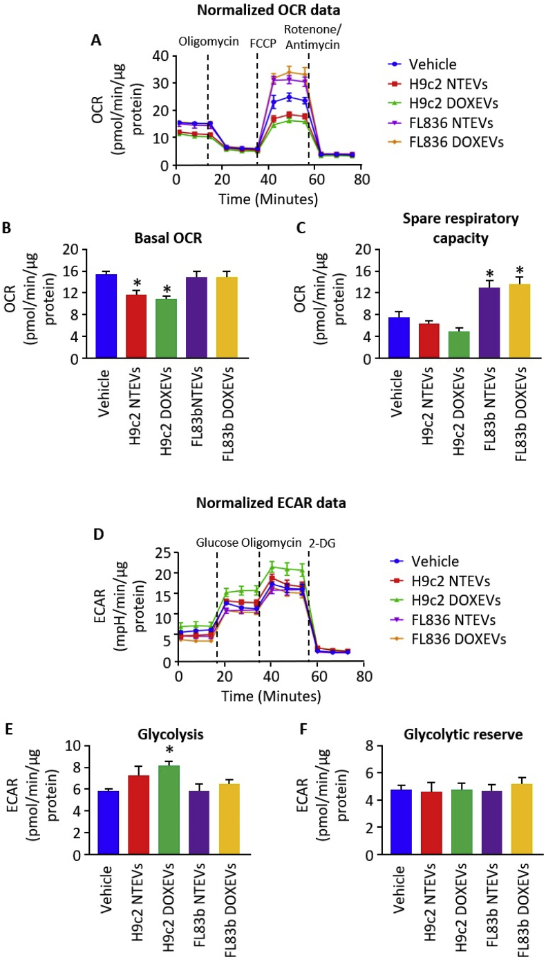
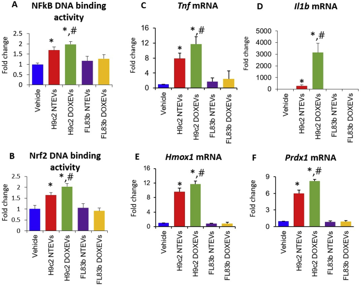
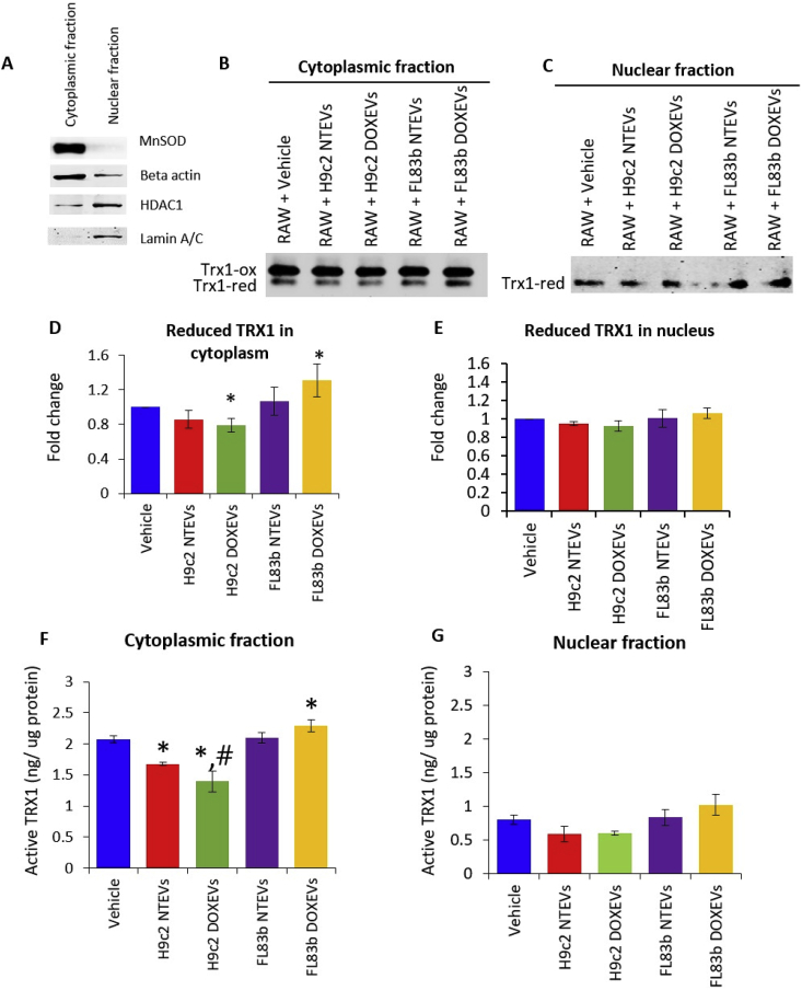
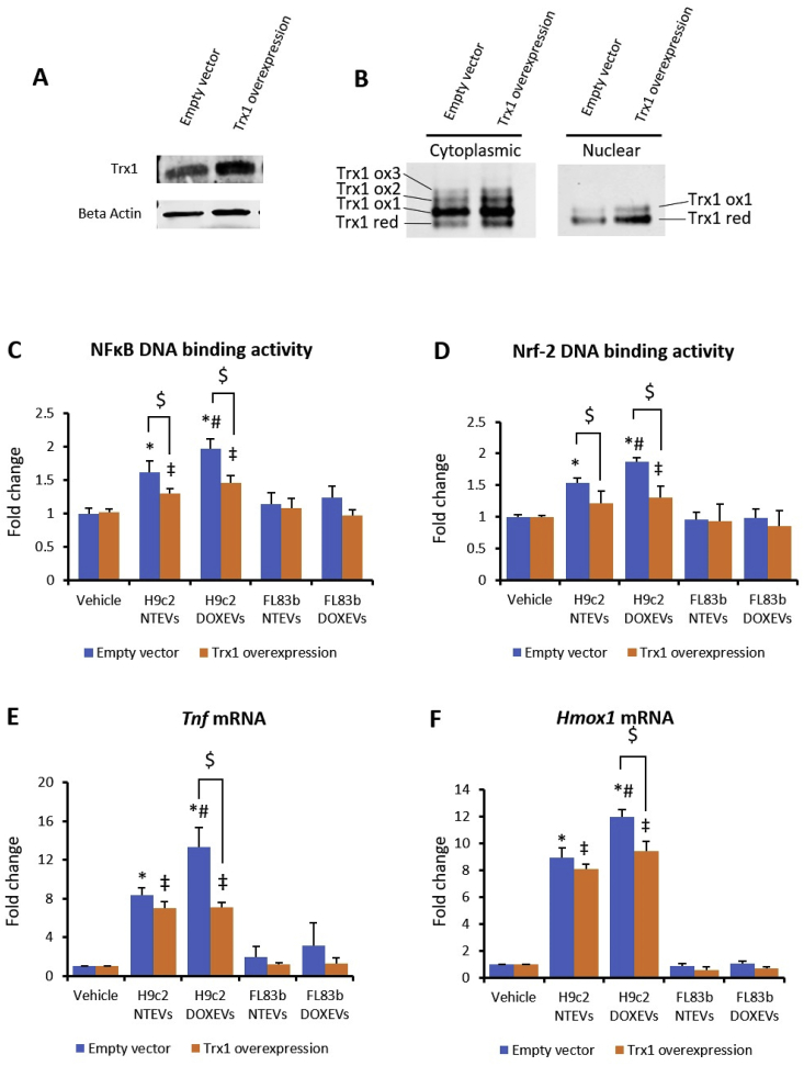
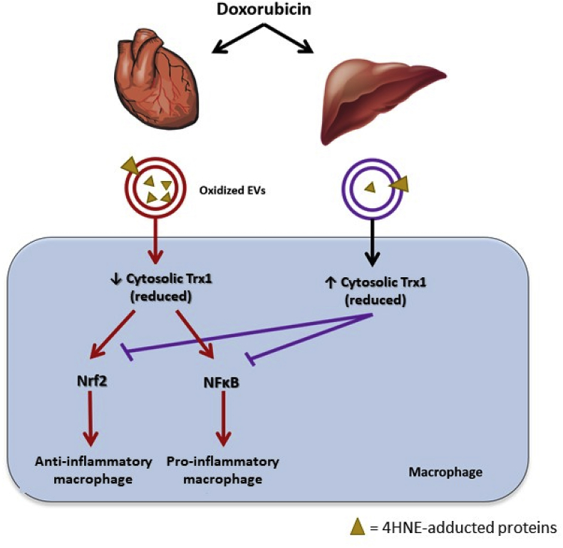
Extracellular vesicles (EVs) generated from redox active anticancer drugs are released into the extracellular environment. These EVs contain oxidized molecules and trigger inflammatory responses by macrophages. Using a mouse model of doxorubicin (DOX)-induced tissue injury, we previously found that the major sources of circulating EVs are from heart and liver, organs that are differentially affected by DOX. Here, we investigated the effects of EVs from cardiomyocytes and those from hepatocytes on macrophage activation. EVs from H9c2 rat cardiomyocytes (H9c2 EVs) and EVs from FL83b mouse hepatocytes (FL83 b EVs) have different levels of protein-bound 4-hydroxynonenal and thus different immunostimulatory effects on mouse RAW264.7 macrophages. H9c2 EVs but not FL83 b EVs induced both pro-inflammatory and anti-inflammatory macrophage activation, mediated by NFκB and Nrf-2 pathways, respectively. DOX enhanced the effects of H9c2 EVs but not FL83 b EVs. While EVs from DOX-treated H9c2 cells (H9c2 DOXEVs) suppressed mitochondrial respiration and increased glycolysis of macrophages, EVs from DOX-treated FL83b cells (FL83b DOXEVs) enhanced mitochondrial reserve capacity. Mechanistically, the different immunostimulatory functions of H9c2 EVs and FL83 b EVs are regulated, in part, by the redox status of the cytoplasmic thioredoxin 1 (Trx1) of macrophages. H9c2 DOXEVs lowered the level of reduced Trx1 in cytoplasm while FL83b DOXEVs did the opposite. Trx1 overexpression alleviated the effect of H9c2 DOXEVs on NFκB and Nrf-2 activation and prevented the upregulation of their target genes. Our findings identify EVs as a novel Trx1-mediated redox mediator of immune response, which greatly enhances our understanding of innate immune responses during cancer therapy.

Keywords: Extracellular vesicles; Macrophage activation; NFκB; Nrf-2; Thioredoxin 1.


Figures

Fig. 1
Fig. 1
Comparison of DOX toxicity, the levels of EVs and the levels of 4HNE protein adduction in the EVs released from cardiomyocytes and hepatocytes (A) Percentage of surviving H9c2 cells and FL83b cells after 0.5 μM DOX treatment for 24 h (green bars) compared to non-treated control (black bars). (B) Amount of EVs released from one million cells of non-treated H9c2, DOX-treated H9c2, non-treated FL83b, or DOX-treated FL83b cells. (C) Western blots of EVs markers: Alix and CD81 in EVs derived from non-treated H9c2 (H9c2 NTEVs), DOX-treated H9c2 (H9c2 DOXEVs), non-treated FL83b (FL83b NTEVs), or DOX-treated FL83b (FL83b DOXEVs). (D) The levels of 4HNE protein adduction in H9c2 NTEVs, H9c2 DOXEVs, FL83b NTEVs, or FL83b DOXEVs. Data represent the mean of band intensities relative to H9c2 NTEVs ±SD of 5 independent experiments * p < 0.05. (For interpretation of the references to color in this figure legend, the reader is referred to the Web version of this article.)
Fig. 2
Fig. 2
Cardiomyocyte-derived EVs but not hepatocyte-derived EVs promote both pro-inflammatory and anti-inflammatory cytokine release as well as nitric oxide production by macrophages. The effect was potentiated by DOX treatment (A-C) Levels of pro-inflammatory cytokines TNF alpha, IL-1 beta, IL-6 in RAW264.7 cells treated for 24 h with vehicle, H9c2 NTEVs, H9c2 DOXEVs, FL83b NTEVs, or FL83b DOXEVs. (D) Level of Nitrite/Nitrate as a parameter of •NO production in RAW264.7 cells treated with vehicle, H9c2 NTEVs, H9c2 DOXEVs, FL83b NTEVs, or FL83b DOXEVs. l-NAME was used as a negative control as it is the inhibitor of •NO production. (EF) Levels of anti-inflammatory cytokines; IL-10 and TGF beta in RAW264.7 cells treated for 24 h with vehicle, H9c2 NTEVs, H9c2 DOXEVs, FL83b NTEVs, or FL83b DOXEVs. Data represent the mean ± SD of 4 independent experiments, *p < 0.05 vs vehicle control, #p < 0.05 vs H9c2 NTEVs. (GI) Gene expression of NR8383 rat macrophages after EVs treatment. Bar graphs represent fold change of Il1b(G), Nos2(H) and Il10(I) gene expression measured by RT-PCR of NR8383 rat macrophages treated with vehicle, H9c2 NTEVs, H9c2 DOXEVs, FL83b NTEVs, or FL83b DOXEVs. Data represent the mean ± SD of 3 independent experiments, *p < 0.05 vs vehicle, #p < 0.05 vs H9c2 NTEVs.
Fig. 3
Fig. 3
Differential effects of H9c2 EVs and FL83b EVs on mitochondrial respiration and glycolysis (A) Kinetic graph of OCR at baseline and after oligomycin, FCCP, and rotenone/antimycin treatment of macrophages incubated with vehicle, H9c2 NTEVs, H9c2 DOXEVs, FL83b NTEVs, or FL83b DOXEVs. (B) Bar graph represents the average of basal OCR calculated from graph A. (C) Bar graph represents the average of Spare Respiratory Capacity calculated from graph A. (D) Kinetic graph of ECAR at baseline and after glucose, oligomycin, and 2-deoxyglucose (2-DG) treatment of macrophages incubated with vehicle, H9c2 NTEVs, H9c2 DOXEVs, FL83b NTEVs, or FL83b DOXEVs. (E) Bar graph represents the average of basal glycolysis calculated from graph D. (F) Bar graph represents the average of glycolytic reserve calculated from graph D. Data represent the mean ± SEM of 3 independent experiments, *p < 0.05 vs vehicle.
Fig. 4
Fig. 4
Cardiomyocyte-derived EVs but not hepatocyte-derived EVs enhanced NFκB and Nrf-2 transcription factor activities. The effect was potentiated by DOX (A) NFkB DNA binding activity and (B) Nrf2 DNA binding activity in macrophages treated with vehicle, H9c2 NTEVs, H9c2 DOXEVs, FL83b NTEVs, or FL83b DOXEVs. (C, D) Fold change of pro-inflammatory cytokines TNF alpha and IL-1 beta gene expression. (E, F) Fold change of anti-oxidant enzymes Hmox1 and Prdx1 gene expression after 24 h treatment of RAW264.7 cells with vehicle, H9c2 NTEVs, H9c2 DOXEVs, FL83b NTEVs, or FL83b DOXEVs. Data represent the mean ± SD of 3 independent experiments, *p < 0.05 vs vehicle, #p < 0.05 vs H9c2 NTEVs.
Fig. 5
Fig. 5
Differential effects of H9c2 EVs and FL83b EVs on reduced Trx1 and Trx1 activity (A) Verification of cytoplasmic and nuclear fraction purification by Western blot of manganese superoxide dismutase (MnSOD) and beta actin for cytoplasmic fraction, as well as histone deacetylase 1 (HDAC1) and lamin A/C for nuclear fraction. (B and C) Redox western of Trx1 in cytoplasmic fraction (B) and nuclear fraction (C) of RAW264.7 treated with vehicle, H9c2 NTEVs, H9c2 DOXEVs, FL83b NTEVs, or FL83b DOXEVs. (D and E) Bar graph shows the average fold change (n = 3) of reduced Trx1 levels in cytoplasm (D) and nuclei (E). (F and G) Trx1 activity from equal amounts of protein in cytoplasm (F) and nuclei (G) of RAW264.7 treated with vehicle, H9c2 NTEVs, H9c2 DOXEVs, FL83b NTEVs, or FL83b DOXEVs, *p < 0.05 vs vehicle, #p < 0.05 vs H9c2 NTEVs.
Fig. 6
Fig. 6
Trx1 overexpression suppressed the effect of H9c2 EVs on macrophage activation via NFκB and Nrf-2 (A) Western blot of Trx1 in RAW264.7 macrophage with Trx1 overexpression compared to empty vector. (B) Redox western of Trx1 in cytoplasmic and nuclear fraction of RAW264.7 macrophage with Trx1 overexpression compared to empty vector. (C and D) NFkB DNA binding activity (C) and Nrf2 DNA binding activity (D) in empty vector (blue) and Trx1 overexpressed RAW264.7 macrophages (orange) treated with vehicle, H9c2 NTEVs, H9c2 DOXEVs, FL83b NTEVs, or FL83b DOXEVs. (E and F) Fold change of pro-inflammatory cytokines Tnf(C) anti-oxidant Hmox1(D) gene expression after 24 h treatment of empty vector or Trx1 overexpressed RAW264.7 cells treated with vehicle, H9c2 NTEVs, H9c2 DOXEVs, FL83b NTEVs, or FL83b DOXEVs. Data represent the mean ± SD of 3 independent experiments, *p < 0.05 vs empty vector + vehicle, #p < 0.05 vs empty vector + H9c2 NTEVs, ‡p < 0.05 vs Trx1 overexpression + vehicle, $ p < 0.05 vs empty vector with the same treatment. (For interpretation of the references to color in this figure legend, the reader is referred to the Web version of this article.)
Fig. 7
Fig. 7
Proposed mechanistic model for the differential effects of normal tissue-derived EVs on macrophages. Cardiomyocyte EVs induce pro- and anti-inflammatory macrophage activation by transferring 4HNE-adducted proteins to the macrophages, which in turn exacerbates oxidative stress in the macrophages by decreasing cytosolic reduced Trx1. As a result of reduced Trx1 exhaustion, NFκB is activated and pro-inflammatory cytokines are produced. Nrf-2 activation occurs as Keap-1 becomes more oxidized by the loss of the Trx1 that is required for it to maintain its reducing state. DOX aggravates the propagation of oxidative stress induced by cardiomyocyte EVs. In contrast, hepatocyte EVs upregulate cytosolic Trx1, which maintains macrophages in an inactivated status. DOX potentiates the effect of hepatocyte EVs on Trx1 induction, thus preventing macrophage activation after Dox treatment.

References

    1. Benedikter B.J., Weseler A.R., Wouters E.F.M., Savelkoul P.H.M., Rohde G.G.U., Stassen F.R.M. Redox-dependent thiol modifications: implications for the release of extracellular vesicles. Cell. Mol. Life Sci. : CM. 2018;75:2321–2337. - PMC - PubMed
    1. Robbins P.D. Extracellular vesicles and aging. Stem Cell Invest. 2017;4:98. - PMC - PubMed
    1. Minciacchi V.R., Freeman M.R., Di Vizio D. Extracellular vesicles in cancer: exosomes, microvesicles and the emerging role of large oncosomes. Semin. Cell Dev. Biol. 2015;40:41–51. - PMC - PubMed
    1. Loyer X., Vion A.C., Tedgui A., Boulanger C.M. Microvesicles as cell-cell messengers in cardiovascular diseases. Circ. Res. 2014;114:345–353. - PubMed
    1. Fujita Y., Kosaka N., Araya J., Kuwano K., Ochiya T. Extracellular vesicles in lung microenvironment and pathogenesis. Trends Mol. Med. 2015;21:533–542. - PubMed

Publication types

MeSH terms

LinkOut - more resources