. 2019 Jul 3;17(7):392.
doi: 10.3390/md17070392.

Enzyme-Aided Extraction of Fucoidan by AMG Augments the Functionality of EPCs through Regulation of the AKT/Rheb Signaling Pathway

Affiliations

Enzyme-Aided Extraction of Fucoidan by AMG Augments the Functionality of EPCs through Regulation of the AKT/Rheb Signaling Pathway

Vinoth Kumar Rethineswaran et al. Mar Drugs. .

Abstract

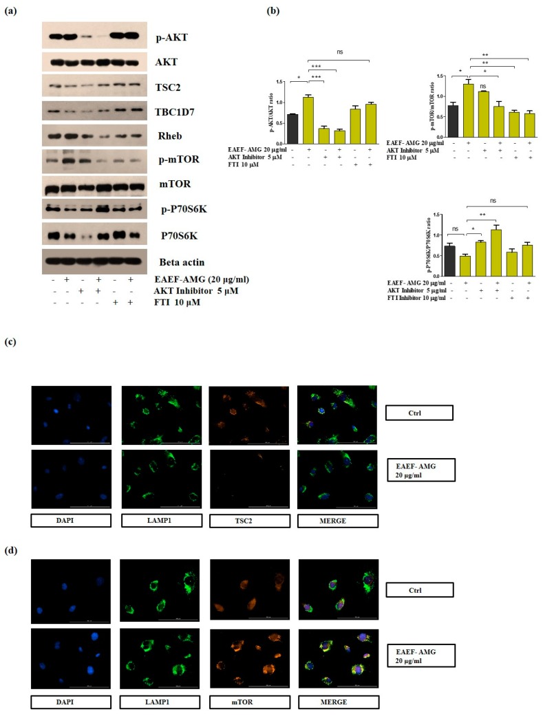
The purpose of the present study is to improve the endothelial progenitor cells (EPC) activation, proliferation, and angiogenesis using enzyme-aided extraction of fucoidan by amyloglucosidase (EAEF-AMG). Enzyme-aided extraction of fucoidan by AMG (EAEF-AMG) significantly increased EPC proliferation by reducing the reactive oxygen species (ROS) and decreasing apoptosis. Notably, EAEF-AMG treated EPCs repressed the colocalization of TSC2/LAMP1 and promoted perinuclear localization of mTOR/LAMP1 and mTOR/Rheb. Moreover, EAEF-AMG enhanced EPC functionalities, including tube formation, cell migration, and wound healing via regulation of AKT/Rheb signaling. Our data provided cell priming protocols to enhance therapeutic applications of EPCs using bioactive compounds for the treatment of CVD.

Keywords: amyloglucosidase; cell proliferation; endothelial progenitor cells; fucoidan; vascular regeneration.


Conflict of interest statement

The authors declare no conflict of interest.

Figures

Figure 1
Figure 1
Enzyme-aided extraction of fucoidan enhances EPCs proliferation and tube formation. (a) Proposed experimental design. (b) Proliferation rate of the EPC cells treated with enzyme-aided extraction of fucoidan treated with equivalent concentration (20 μg/mL) of AMG, viscozyme, or pectinex for 24 h assessed using WTS-1. (ce) Tube formation assay was performed using matrigel coated plates for 6 h, then capillary structure was visualized using a light microscopy (Olympus, Tokyo, Japan), total tube length and branches were quantified using ImageJ software (NIH, Bethesda, MD, USA). Data are presented as mean ± standard error of the mean (SEM). The results are considered statistically significant at * p < 0.05; ** p < 0.01; *** p < 0.001 when compared to untreated groups.
Figure 2
Figure 2
EAEF-AMG mitigates apoptotic cell death and enhances angiogenic activity. (a) Cells were treated with EAEF-AMG (10, 20 μg/mL) for 24 and 48 h, then proliferation was measured by WST-1. (b,c) Carboxy-H2DFFDA was used to measure cellular ROS, cells were pretreated with EAEF-AMG (20 μg/mL) for 24 h, followed by H2O2 (250 μM) for 5 min, then ROS was measured by FACS. (d) Apoptotic cells were measured by Annexin V FITC and propidium iodide staining using FACS. (e) For tube formation assay, cells were treated with EAEF-AMG (20 μg/mL) for 6 h, following which capillary structures were visualized using a light microscope (Olympus, Tokyo, Japan). Total tube length and branches were quantified using ImageJ software (NIH, Bethesda, MD, USA). (f) Scratch wound healing assays, were performed by cell scratcher for 6 h. Wound healing area was measured using ImageJ software (NIH, Bethesda, MD, USA). (g) Transwell migration was performed by seeding cells in the upper inserts of the transwell chamber, whereas medium with EAEF-AMG was added to the lower chamber. The cells were incubated for 6 h and the number of migrated cells was counted in three random fields for each filter (magnification, 20x) under a microscope. Data are presented as mean ± standard error of the mean (SEM). The results are considered as statistically significant at * p < 0.05; **p < 0.01; ***p < 0.001 when compared to untreated groups.
Figure 2
Figure 2
EAEF-AMG mitigates apoptotic cell death and enhances angiogenic activity. (a) Cells were treated with EAEF-AMG (10, 20 μg/mL) for 24 and 48 h, then proliferation was measured by WST-1. (b,c) Carboxy-H2DFFDA was used to measure cellular ROS, cells were pretreated with EAEF-AMG (20 μg/mL) for 24 h, followed by H2O2 (250 μM) for 5 min, then ROS was measured by FACS. (d) Apoptotic cells were measured by Annexin V FITC and propidium iodide staining using FACS. (e) For tube formation assay, cells were treated with EAEF-AMG (20 μg/mL) for 6 h, following which capillary structures were visualized using a light microscope (Olympus, Tokyo, Japan). Total tube length and branches were quantified using ImageJ software (NIH, Bethesda, MD, USA). (f) Scratch wound healing assays, were performed by cell scratcher for 6 h. Wound healing area was measured using ImageJ software (NIH, Bethesda, MD, USA). (g) Transwell migration was performed by seeding cells in the upper inserts of the transwell chamber, whereas medium with EAEF-AMG was added to the lower chamber. The cells were incubated for 6 h and the number of migrated cells was counted in three random fields for each filter (magnification, 20x) under a microscope. Data are presented as mean ± standard error of the mean (SEM). The results are considered as statistically significant at * p < 0.05; **p < 0.01; ***p < 0.001 when compared to untreated groups.
Figure 3
Figure 3
EAEF-AMG enhanced the functional markers expression of EPCs. Cells treated with EAEF-AMG. (20 μg/mL) for 24 h showed increased expression of CD34, CXCR4, C-Kit, VEGFR2, and VE-Cadherin using fluorescence activated cell sorting (FACS). FACS gating was performed using non-stained cells as a negative control. The fraction of positively stained cells was determined by comparison with non-stained cells. The percentage of positively-stained cells is indicated by the positive peaks (red lines indicate cells stained with each antibodies, and black lines indicate the negative control cells).
Figure 4
Figure 4
EAEF-AMG regulates the AKT/Rheb signaling pathway. (a) Cells were treated with EAEF-AMG (20 μg/mL) and in combination with specific inhibitors such as AKT inhibitor (5 μM), and farnesyltransferase inhibitor (10 μM) for 24 h, then Western blots were performed to evaluate the protein level expression of p-AKT, AKT, TSC2, and TBCD17, Rheb, p-mTOR, mTOR, p-P70S6K, and P70S6K and loading control beta actin. (b) Quantification of Western blots. (cd) Immunofluorescence was performed by treating cells with EAEF-AMG (20 μg/mL) for 45 min to assess the localization of TSC2/LAMP1 in perinuclear sites, and images of perinuclear aggregation of mTOR/LAMP1 were captured using a 40× objective lens on a Lion Heart FX automated microscope (Biotek, Winooski, VT, USA). Scale bar = 100 μm.
Figure 5
Figure 5
EAEF-AMG enhances angiogenic activity via regulation of AKT/Rheb signaling. (a,b) Cells treated with EAEF-AMG (20 μg/mL) and in combination with AKT inhibitor (5 μM) and farnesyltransferase inhibitor (10 μM) for 24 h, then proliferation was measured by WST-1. (c) Scratch wound healing assay was performed using a cell scratcher and wound healing area was measured after 6 h using ImageJ software (NIH, Bethesda, MD, USA). (d) Transwell migration was performed by seeding cells on upper inserts and the lower chamber selectively contained EAEF-AMG, AKT, and farnesyltransferase inhibitor. Next, cells were incubated for 6 h and the number of migrated cells was counted in three random fields per filter (magnification, 20×) using a light microscope. (eg) In the tube formation assay, capillary structures were captured after 6 h using a light microscope (Olympus, Tokyo, Japan). Total tube length and branches were quantified using ImageJ software (NIH, Bethesda, MD, USA). Data are presented as mean ± standard error of the mean (SEM). The results are considered as statistically significant at * p < 0.05; ** p < 0.01; *** p < 0.001 when compared to untreated groups.
Figure 6
Figure 6
Schematic diagram representation of EAEF-AMG improving angiogenic activity through regulation of AKT/Rheb signaling. Exposure of EAEF-AMG up-regulated the expression of p-AKT, leading to obstruction of the perinuclear accumulation and colocalization of TSC2/LAMP1, followed by augmenting the perinuclear aggregation of mTOR/Lamp1, which further regulates downstream signaling, and intensifies EPC functionality.

Similar articles

Cited by

References

    1. Kivimaki M., Steptoe A. Effects of stress on the development and progression of cardiovascular disease. Nat. Rev. Cardiol. 2018;15:215–229. doi: 10.1038/nrcardio.2017.189. - DOI - PubMed
    1. Duncker D.J., Koller A., Merkus D., Canty J.M., Jr. Regulation of coronary blood flow in health and ischemic heart disease. Prog. Cardiovasc. Dis. 2015;57:409–422. doi: 10.1016/j.pcad.2014.12.002. - DOI - PMC - PubMed
    1. Yu P., Zhang Z., Li S., Wen X., Quan W., Tian Q., Chen J., Zhang J., Jiang R. Progesterone modulates endothelial progenitor cell (EPC) viability through the CXCL12/CXCR4/PI3K/Akt signalling pathway. Cell Prolif. 2016;49:48–57. doi: 10.1111/cpr.12231. - DOI - PMC - PubMed
    1. Masuda H., Alev C., Akimaru H., Ito R., Shizuno T., Kobori M., Horii M., Ishihara T., Isobe K., Isozaki M., et al. Methodological development of a clonogenic assay to determine endothelial progenitor cell potential. Circ. Res. 2011;109:20–37. doi: 10.1161/CIRCRESAHA.110.231837. - DOI - PubMed
    1. Ke X., Shu X.R., Wu F., Hu Q.S., Deng B.Q., Wang J.F., Nie R.Q. Overexpression of the beta2AR gene improves function and re-endothelialization capacity of EPCs after arterial injury in nude mice. Stem Cell Res. Ther. 2016;7:73. doi: 10.1186/s13287-016-0335-y. - DOI - PMC - PubMed

MeSH terms

Substances

LinkOut - more resources