. 2019 Jul 3;11(7):604.
doi: 10.3390/v11070604.

Anti-Respiratory Syncytial Virus Activity of Plantago asiatica and Clerodendrum trichotomum Extracts In Vitro and In Vivo

Affiliations

Anti-Respiratory Syncytial Virus Activity of Plantago asiatica and Clerodendrum trichotomum Extracts In Vitro and In Vivo

Kiramage Chathuranga et al. Viruses. .

Abstract

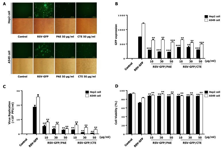
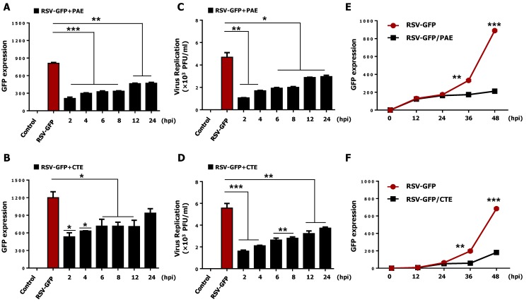
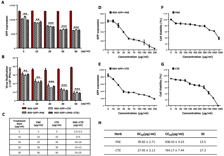
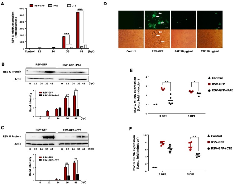
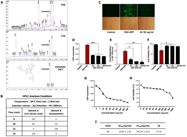
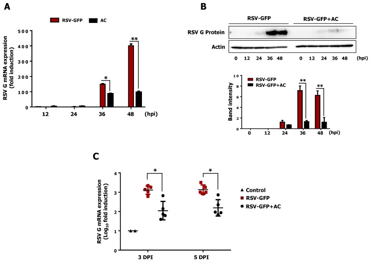
The herbs Plantago asiatica and Clerodendrum trichotomum have been commonly used for centuries in indigenous and folk medicine in tropical and subtropical regions of the world. In this study, we show that extracts from these herbs have antiviral effects against the respiratory syncytial virus (RSV) in vitro cell cultures and an in vivo mouse model. Treatment of HEp2 cells and A549 cells with a non-cytotoxic concentration of Plantago asiatica or Clerodendrum trichotomum extract significantly reduced RSV replication, RSV-induced cell death, RSV gene transcription, RSV protein synthesis, and also blocked syncytia formation. Interestingly, oral inoculation with each herb extract significantly improved viral clearance in the lungs of BALB/c mice. Based on reported information and a high-performance liquid chromatography (HPLC) analysis, the phenolic glycoside acteoside was identified as an active chemical component of both herb extracts. An effective dose of acteoside exhibited similar antiviral effects as each herb extract against RSV in vitro and in vivo. Collectively, these results suggest that extracts of Plantago asiatica and Clerodendrum trichotomum could provide a potent natural source of an antiviral drug candidate against RSV infection.

Keywords: Clerodendrum trichotomum; Plantago asiatica; RSV; acteoside; therapeutic effects.


Conflict of interest statement

The authors declare no conflict of interest.

Figures

Figure 1
Figure 1
Antiviral activity of Plantago asiatica extract (PAE) and Clerodendrum trichotomum extract (CTE) in HEp2 cells and A549 cells. HEp2 cells and A549 cells were seeded into 12 well cell culture plates with the cell number of 2.5 × 10⁵ cells/well. Twelve hours later, the medium was changed to 1% fetal bovine serum (FBS) containing Dulbecco’s Modified Eagle’s Medium (DMEM) and cells were infected with Green Fluorescent Protein fused Respiratory syncytial virus (RSV-GFP) 0.1multiplicity of infection (MOI) or kept uninfected. Two hours later, the medium was replaced with 10% FBS containing DMEM and cells were treated with 10, 30, 50 (μg/mL) PAE or CTE. Cells without any treatment regard as virus only. (A) After 48 h, images were obtained (200× magnification). (B) GFP absorbance levels were measured by Gloma multi-detection luminometer (Promega). (C) Virus titration was done from the cell supernatant and cells by standard plaque assay and expressed as plaque forming unit (PFU). (D) Cell viability was determined by trypan blue exclusion assay at 48hour post infection (hpi). GFP absorbance, cell viability and virus titer expressed as mean ± standard deviations (SD). Error bars indicate the range of values obtained from counting duplicate in three independent experiments (** p < 0.01 and *** p < 0.001 regarded as significant difference).
Figure 2
Figure 2
The therapeutic effect of PAE and CTE against RSV-GFP infection. HEp2 cells were seeded into 12 well cell culture plates and left for 12 h. Medium was changed with DMEM containing 1% FBS and cells were infected with RSV-GFP (0.1MOI) for 2 h. (A,B) RSV-GFP infected cells were treated with 50 μg/mL PAE or CTE at different times after post infection as indicated or left untreated, and GFP expression level was measured at 48 hpi. (C,D) Virus titer was measured from both supernatant and cells by standard plaque assay at 48 hpi and expressed as PFU. (E,F) Cells were treated with 50 μg/mL PAE or CTE, and GFP expression level was measured at different time after virus infection as indicated. GFP absorbance and virus titer expressed as mean ± SD. Error bars indicate the range of values obtained from counting duplicate in three independent experiments (* p < 0.05, ** p < 0.01 and *** p < 0.001 regarded as significant difference).
Figure 3
Figure 3
Synergistic effect and effective concentration (EC50), cytotoxic concentration (CC50) of PAE and CTE in HEp2 cells. (A) HEp2 cells were infected with RSV-GFP (0.1 MOI) for 2 h with DMEM containing 1% FBS. Cells were treated with PAE, CTE or combination of both at different concentrations with DMEM containing 10% FBS. At 48 hpi GFP expression level was determined. (B) Virus titer was measured from both supernatant and cells by standard plaque assay at 48 hpi and expressed as PFU. (C) Dose information of PAE and CTE single or combination treatment. (D,E) HEp2 cells were infected with RSV-GFP (O.1MOI) for 2 h with DMEM containing 1% FBS. Then, the medium was changed to DMEM containing 10% FBS and cells were treated with various concentrations of PAE (D) or CTE (E). 48 hpi GFP expression level was determined. (F,G) HEp2 cells were treated with various concentrations of PAE (F) or CTE (G), and cell viability was determined at 48 h post treatment (hpt) by cell cytotoxicity assay kit. (H) To calculate EC50 value, 50% reduction of GFP expression was considered as equivalent to the 50% reduction in virus titer. The ratio between CC50 and EC50 was considered as Selectivity Index (SI). GFP absorbance and cell viability expressed as mean ± SD. Error bars indicate the range of values obtained from counting duplicate in three independent experiments. (** p < 0.01 and *** p < 0.001 regarded as significant difference).
Figure 4
Figure 4
Reduction of RSV Glycoprotein (RSV-G) gene transcription, protein translation and syncytium formation in-vitro and inhibition of RSV replication in-vivo by PAE and CTE. HEp2 cells were seeded in six well cell culture plats and incubate for 12 h. Medium was changed into DMEM containing 1% FBS and RSV-GFP (0.1MOI) was infected for 2 h. Then cells were treated with 50 μg/mL PAE or CTE. (A) Cells were harvested at indicated time points, and RSV-G protein mRNA level was measured at 12, 24, 36, 48 hpi by qRT-PCR, Glyceraldehyde 3-phosphate dehydrogenase (GAPDH) was used for the normalization. (B,C) Immunoblot analysis was performed using cell lysates harvested at indicated time points to measure RSV-G protein and β-actin protein expression level time-dependently. The intensity of the RSV-G was quantified. (D) Cell and the GFP image were taken at 48 hpi to see the syncytial formation inhibition by PAE and CTE (400× magnification). (E,F) Five weeks old (16 g/mice) BALB/c mice (n = 5) were intranasally infected with RSV-GFP (1 × 10⁶ PFU/mice) in the total volume of 28 μL. PAE or CTE were orally administrated at a dose of 200 μL/mice (0.5 mg/mL) at 6, 12, 18 and 24 hpi. At 3 and 5-day post infection (dpi), lung tissues were collected, and the transcription level of RSV-G protein mRNA was determined by qRT-PCR. The arrow indicates the RSV syncytium formation in HEp2 cells. mRNA expression, band intensity expressed as mean ± SD. Error bars indicate the range of values obtained from three independent experiments. In vivo experiment was performed in duplicate. (* p < 0.05, ** p < 0.01 and *** p < 0.001 regarded as significant difference).
Figure 5
Figure 5
Identification and antiviral effect of acteoside (AC) in-vitro. (A,B) Chemical compounds in PAE and CTE were analyzed by the reversed phase HPLC. The monolayer of HEp2 cells was infected with RSV-GFP (0.1MOI) for 2 h with DMEM containing 1% FBS. Then, the medium was replaced with DMEM containing 10% FBS and cells were treated with 10, 30, 50 (ng/mL) AC. (C) After 48 h, images were obtained (200× magnification). (D) GFP absorbance levels were measured by Gloma multi-detection luminometer (Promega). (E) Viruses were titrated from the cell supernatant and cells by standard plaque assay. (F) Cell viability was determined by trypan blue exclusion assay at 48 hpi. (A) HEp2 cells were infected with RSV-GFP (O.1MOI) for 2 h with DMEM containing 1% FBS. Then, the medium was changed to DMEM containing 10% FBS and cells were treated with various concentrations AC. 48 hpi GFP expression level was determined. (G) HEp2 cells were treated with various concentrations of AC and cell viability was determined at 48 hpi by cell cytotoxicity assay kit. (H) To calculate EC50 value, 50% reduction of GFP expression was considered as equivalent to the 50% reduction in virus titer. (I) The ratio between CC50 and EC50 considered as Selectivity Index (SI). GFP absorbance, cell viability and virus titer expressed as mean ± SD. Error bars indicate the range of values obtained from counting duplicate in three independent experiments. In vivo experiment was performed in duplicate. (* p < 0.05, ** p < 0.01 and *** p < 0.001 regarded as significant difference).
Figure 6
Figure 6
Antiviral effect of acteoside in-vitro and in-vivo. (A) RSV-GFP infected cells were treated with 50 ng/mL of AC at 2 hpi and cells were harvested at indicated time points. RSV-G protein mRNA transcription level was determined by qRT-PCR. GAPDH was used for normalization. (B) Infected cells were treated with 50 ng/mL concentration of AC at 2 hpi and cells were harvested at indicated time points. RSV-G protein expression was determined by immunoblotting with anti-RSV-G protein antibody, and the intensity of the RSV-G was quantified. (C) Five weeks old (16 g/mice) BALB/c mice (n = 5) were intranasally infected with RSV-GFP (1 × 10⁶ PFU/mice) in the total volume of 28 μL. 6 hpi AC was intraperitoneally administrated at a dose of 80 mg/Kg body weight of mice. At 3 and 5 dpi, lung tissues were collected, and the transcription level of RSV-G protein mRNA was determined by qRT-PCR. mRNA expression and band intensity expressed as mean ± SD. Error bars indicate the range of values obtained from three independent experiments. In vivo experiment was performed in duplicate. (* p < 0.05 and ** p < 0.01 regarded as significant difference).

Similar articles

Cited by

References

    1. Piedimonte G., Perez M.K. Alternative Mechanisms for Respiratory Syncytial Virus (RSV) Infection and Persistence: Could RSV Be Transmitted Through the Placenta And Persist Into Developing Fetal Lungs? Curr. Opin. Pharmacol. 2014;16:82–88. doi: 10.1016/j.coph.2014.03.008. - DOI - PMC - PubMed
    1. Mazur N.I., Martinón-Torres F., Baraldi E., Fauroux B., Greenough A., Heikkinen T., Manzoni P., Mejias A., Nair H., Papadopoulos N.G., et al. Lower Respiratory Tract Infection Caused by Respiratory Syncytial Virus: Current Management and New Therapeutics. Lancet Respir. Med. 2015;3:888–900. doi: 10.1016/S2213-2600(15)00255-6. - DOI - PubMed
    1. Walsh E., Falsey A.R. Respiratory Syncytial Virus Infection in Adult Populations. Infect. Disord.-Drug Targets. 2012;12:98–102. doi: 10.2174/187152612800100116. - DOI - PubMed
    1. Le Nouen C., Brock L.G., Luongo C., McCarty T., Yang L., Mehedi M., Wimmer E., Mueller S., Collins P.L., Buchholz U.J., et al. Attenuation of Human Respiratory Syncytial Virus by Genome-Scale Codon-Pair Deoptimization. Proc. Natl. Acad. Sci. USA. 2014;111:13169–13174. doi: 10.1073/pnas.1411290111. - DOI - PMC - PubMed
    1. Kneyber M.C., Moll H.A., De Groot R. Treatment and prevention of respiratory syncytial virus infection. Eur. J. Pediatr. 2000;159:399–411. doi: 10.1007/s004310051296. - DOI - PubMed

Publication types

MeSH terms

LinkOut - more resources