

Marine Morbilliviruses: Diversity and Interaction with Signaling Lymphocyte Activation Molecules
- PMID: 31277275
- PMCID: PMC6669707
- DOI: 10.3390/v11070606
Marine Morbilliviruses: Diversity and Interaction with Signaling Lymphocyte Activation Molecules
Abstract
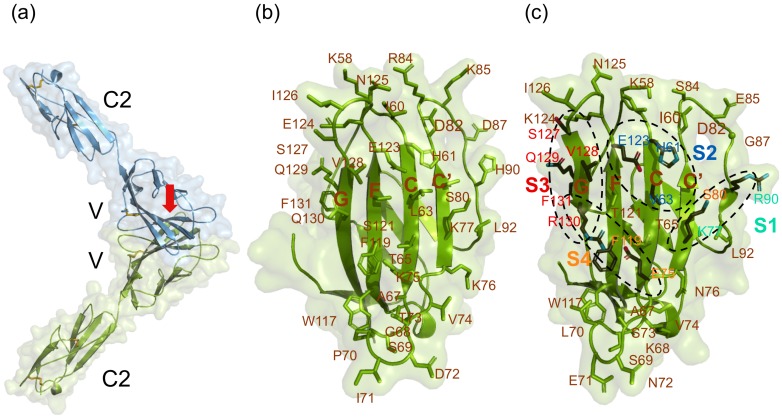
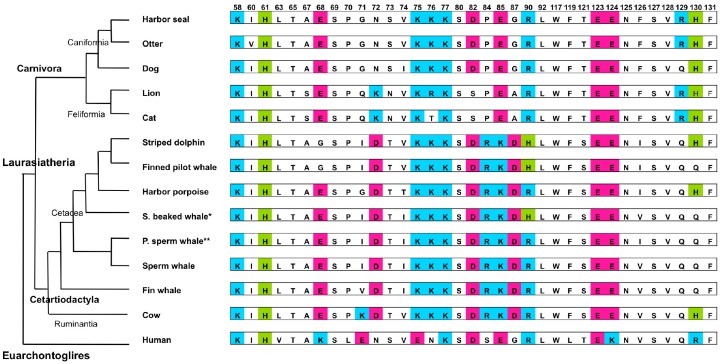
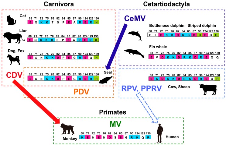
Epidemiological reports of phocine distemper virus (PDV) and cetacean morbillivirus (CeMV) have accumulated since their discovery nearly 30 years ago. In this review, we focus on the interaction between these marine morbilliviruses and their major cellular receptor, the signaling lymphocyte activation molecule (SLAM). The three-dimensional crystal structure and homology models of SLAMs have demonstrated that 35 residues are important for binding to the morbillivirus hemagglutinin (H) protein and contribute to viral tropism. These 35 residues are essentially conserved among pinnipeds and highly conserved among the Caniformia, suggesting that PDV can infect these animals, but are less conserved among cetaceans. Because CeMV can infect various cetacean species, including toothed and baleen whales, the CeMV-H protein is postulated to have broader specificity to accommodate more divergent SLAM interfaces and may enable the virus to infect seals. In silico analysis of viral H protein and SLAM indicates that each residue of the H protein interacts with multiple residues of SLAM and vice versa. The integration of epidemiological, virological, structural, and computational studies should provide deeper insight into host specificity and switching of marine morbilliviruses.
Keywords: cetacean morbillivirus; host specificity; marine mammal; morbillivirus; phocine distemper virus; receptor; signaling lymphocyte activation molecule.
Conflict of interest statement
The authors declare no conflicts of interest.
Figures
Similar articles
-
Cetacean morbillivirus: current knowledge and future directions.Viruses. 2014 Dec 22;6(12):5145-81. doi: 10.3390/v6125145. Viruses. 2014. PMID: 25533660 Free PMC article. Review.
-
Canine and Phocine Distemper Viruses: Global Spread and Genetic Basis of Jumping Species Barriers.Viruses. 2019 Oct 14;11(10):944. doi: 10.3390/v11100944. Viruses. 2019. PMID: 31615092 Free PMC article. Review.
-
Emerging morbillivirus infections of marine mammals: development of two diagnostic approaches.Ann N Y Acad Sci. 2002 Oct;969:51-9. doi: 10.1111/j.1749-6632.2002.tb04350.x. Ann N Y Acad Sci. 2002. PMID: 12381563
-
Host-virus specificity of morbilliviruses predicted by structural modeling of the marine mammal SLAM, a receptor.Comp Immunol Microbiol Infect Dis. 2010 May;33(3):227-41. doi: 10.1016/j.cimid.2008.10.003. Epub 2008 Nov 22. Comp Immunol Microbiol Infect Dis. 2010. PMID: 19027953
-
Phocine distemper virus uses phocine and other animal SLAMs as a receptor but not human SLAM.Microbiol Immunol. 2020 Aug;64(8):578-583. doi: 10.1111/1348-0421.12788. Epub 2020 Jul 25. Microbiol Immunol. 2020. PMID: 32215955
Cited by
-
Molecular fossils illuminate the evolution of retroviruses following a macroevolutionary transition from land to water.PLoS Pathog. 2021 Jul 12;17(7):e1009730. doi: 10.1371/journal.ppat.1009730. eCollection 2021 Jul. PLoS Pathog. 2021. PMID: 34252162 Free PMC article.
-
Identification of Viruses in Molossus Bats from the Brazilian Amazon: A Descriptive Metagenomic Analysis.Microorganisms. 2024 Mar 16;12(3):593. doi: 10.3390/microorganisms12030593. Microorganisms. 2024. PMID: 38543644 Free PMC article.
-
Paramyxoviruses from bats: changes in receptor specificity and their role in host adaptation.Curr Opin Virol. 2023 Feb;58:101292. doi: 10.1016/j.coviro.2022.101292. Epub 2022 Dec 9. Curr Opin Virol. 2023. PMID: 36508860 Free PMC article. Review.
-
Metagenomics-enabled reverse-genetics assembly and characterization of myotis bat morbillivirus.Nat Microbiol. 2023 Jun;8(6):1108-1122. doi: 10.1038/s41564-023-01380-4. Epub 2023 May 4. Nat Microbiol. 2023. PMID: 37142773 Free PMC article.
-
Complete Genome Sequencing of the Divergent Guiana Dolphin Morbillivirus (GDMV), Brazil.Viruses. 2025 Apr 18;17(4):582. doi: 10.3390/v17040582. Viruses. 2025. PMID: 40285024 Free PMC article.
References
-
- Griffin D.E. Measles Virus. In: Knipe D.M., Howley P.M., editors. Fields’ Virology. 3rd ed. Lippincott Williams and Wilkins; New York, NY, USA: 2001. pp. 1401–1442.
Publication types
MeSH terms
Substances
Supplementary concepts
LinkOut - more resources
Full Text Sources
