

Wiring the Binocular Visual Pathways
- PMID: 31277365
- PMCID: PMC6651880
- DOI: 10.3390/ijms20133282
Wiring the Binocular Visual Pathways
Abstract
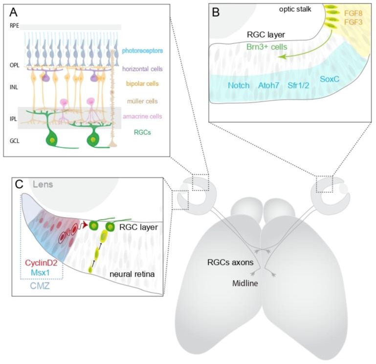
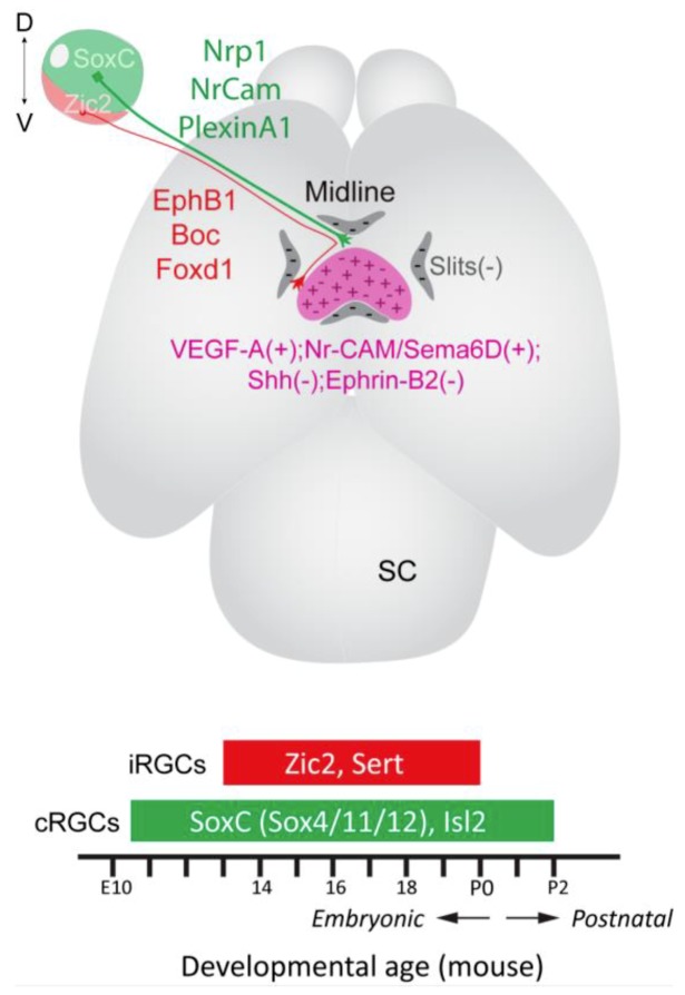
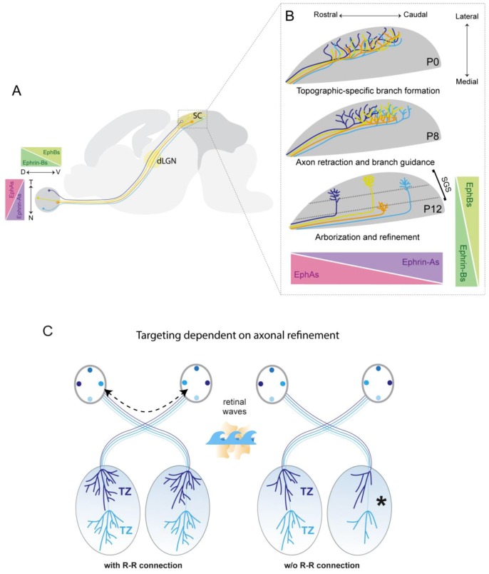
Retinal ganglion cells (RGCs) extend axons out of the retina to transmit visual information to the brain. These connections are established during development through the navigation of RGC axons along a relatively long, stereotypical pathway. RGC axons exit the eye at the optic disc and extend along the optic nerves to the ventral midline of the brain, where the two nerves meet to form the optic chiasm. In animals with binocular vision, the axons face a choice at the optic chiasm-to cross the midline and project to targets on the contralateral side of the brain, or avoid crossing the midline and project to ipsilateral brain targets. Ipsilaterally and contralaterally projecting RGCs originate in disparate regions of the retina that relate to the extent of binocular overlap in the visual field. In humans virtually all RGC axons originating in temporal retina project ipsilaterally, whereas in mice, ipsilaterally projecting RGCs are confined to the peripheral ventrotemporal retina. This review will discuss recent advances in our understanding of the mechanisms regulating specification of ipsilateral versus contralateral RGCs, and the differential guidance of their axons at the optic chiasm. Recent insights into the establishment of congruent topographic maps in both brain hemispheres also will be discussed.
Keywords: axon guidance; neurogenesis; progenitor cell; projection; refinement; retina.
Conflict of interest statement
The authors declare no conflict of interest.
Figures
Similar articles
-
CXCL12 promotes the crossing of retinal ganglion cell axons at the optic chiasm.Development. 2024 Jan 15;151(2):dev202446. doi: 10.1242/dev.202446. Epub 2024 Jan 15. Development. 2024. PMID: 38095299 Free PMC article.
-
The winged helix transcription factor Foxg1 facilitates retinal ganglion cell axon crossing of the ventral midline in the mouse.Development. 2004 Aug;131(15):3773-84. doi: 10.1242/dev.01246. Epub 2004 Jul 7. Development. 2004. PMID: 15240555 Free PMC article.
-
Foxd1 is required for proper formation of the optic chiasm.Development. 2004 Nov;131(22):5727-39. doi: 10.1242/dev.01431. Development. 2004. PMID: 15509772
-
Retinal Ganglion Cell Axon Wiring Establishing the Binocular Circuit.Annu Rev Vis Sci. 2020 Sep 15;6:215-236. doi: 10.1146/annurev-vision-091517-034306. Epub 2020 May 12. Annu Rev Vis Sci. 2020. PMID: 32396770 Review.
-
Development of the Binocular Circuit.Annu Rev Neurosci. 2024 Aug;47(1):303-322. doi: 10.1146/annurev-neuro-111020-093230. Epub 2024 Jul 1. Annu Rev Neurosci. 2024. PMID: 38635868 Review.
Cited by
-
Wiring subcortical image-forming centers: Topography, laminar targeting, and map alignment.Curr Top Dev Biol. 2021;142:283-317. doi: 10.1016/bs.ctdb.2020.10.004. Epub 2020 Nov 16. Curr Top Dev Biol. 2021. PMID: 33706920 Free PMC article. Review.
-
Ophthalmological Manifestations of Oculocutaneous and Ocular Albinism: Current Perspectives.Clin Ophthalmol. 2022 May 24;16:1569-1587. doi: 10.2147/OPTH.S329282. eCollection 2022. Clin Ophthalmol. 2022. PMID: 35637898 Free PMC article. Review.
-
Retinal ganglion cell repopulation for vision restoration in optic neuropathy: a roadmap from the RReSTORe Consortium.Mol Neurodegener. 2023 Sep 21;18(1):64. doi: 10.1186/s13024-023-00655-y. Mol Neurodegener. 2023. PMID: 37735444 Free PMC article. Review.
-
The Slit-Robo signalling pathway in nervous system development: a comparative perspective from vertebrates and invertebrates.Open Biol. 2025 Jul;15(7):250026. doi: 10.1098/rsob.250026. Epub 2025 Jul 9. Open Biol. 2025. PMID: 40628293 Free PMC article. Review.
-
Neurogenesis and Specification of Retinal Ganglion Cells.Int J Mol Sci. 2020 Jan 10;21(2):451. doi: 10.3390/ijms21020451. Int J Mol Sci. 2020. PMID: 31936811 Free PMC article. Review.
