

Assessment of Bandaged Burn Wounds Using Porcine Skin and Millimetric Radiometry
- PMID: 31277437
- PMCID: PMC6651191
- DOI: 10.3390/s19132950
Assessment of Bandaged Burn Wounds Using Porcine Skin and Millimetric Radiometry
Abstract
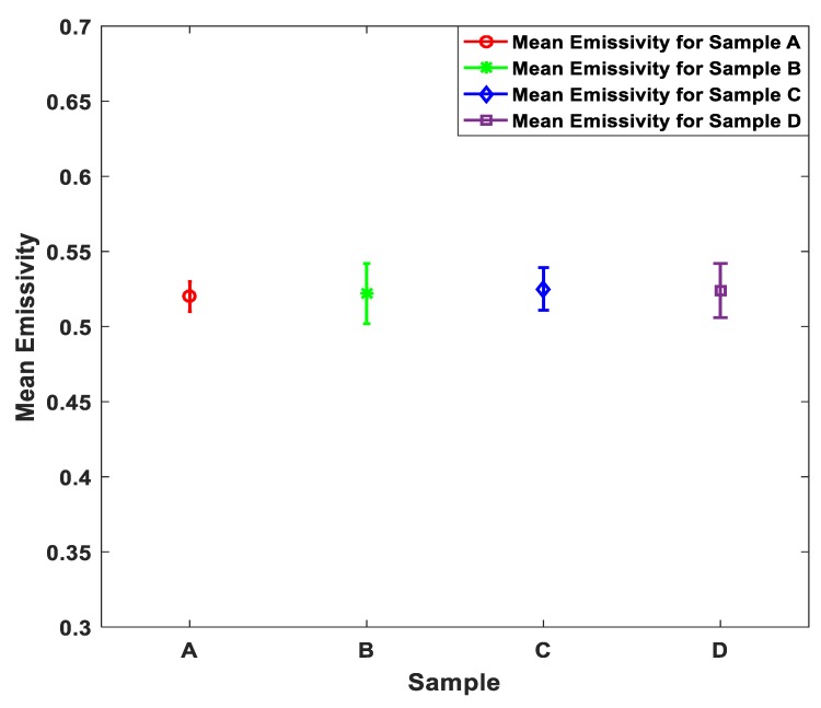
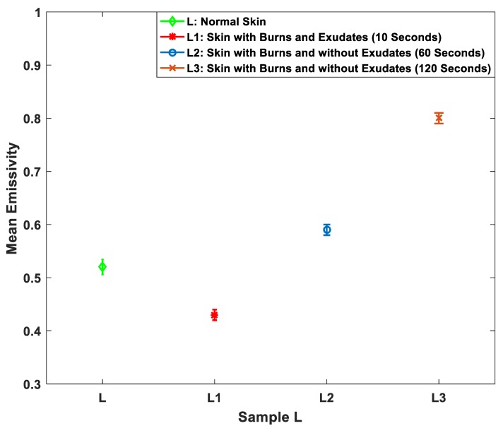
This paper describes the experimental setup and measurements of the emissivity of porcine skin samples over the band of 80-100 GHz. Measurements were conducted on samples with and without dressing materials and before and after the application of localized heat treatments. Experimental measurements indicate that the differences in the mean emissivity values between unburned skin and burned damaged skin was up to ~0.28, with an experimental measurement uncertainty of ±0.005. Measured differences in the mean emissivity values between unburned and burn damaged skin increases with the depth of the burn, indicating a possible non-contact technique for assessing the degree of a burn. The mean emissivity of the dressed burned skin was found to be slightly higher than the undressed burned skin, typically ~0.01 to ~0.02 higher. This indicates that the signature of the burn caused by the application of localized heat treatments is observable through dressing materials. These findings reveal that radiometry, as a non-contact method, is capable of distinguishing between normal and burn-damaged skin under dressing materials without their often-painful removal. This indicates the potential of using millimeter wave (MMW) radiometry as a new type of medical diagnostic to monitor burn wounds.
Keywords: burn wound; dressing materials; millimeter-wave; passive imaging; radiometry.
Conflict of interest statement
The authors declare no conflict of interest.
Figures


Similar articles
-
Synthetic Aperture Radar Imaging for Burn Wounds Diagnostics.Sensors (Basel). 2020 Feb 5;20(3):847. doi: 10.3390/s20030847. Sensors (Basel). 2020. PMID: 32033414 Free PMC article.
-
Passive Millimeter-Wave Imaging for Burns Diagnostics under Dressing Materials.Sensors (Basel). 2022 Mar 22;22(7):2428. doi: 10.3390/s22072428. Sensors (Basel). 2022. PMID: 35408043 Free PMC article.
-
Millimeter-wave emissivity as a metric for the non-contact diagnosis of human skin conditions.Bioelectromagnetics. 2017 Oct;38(7):559-569. doi: 10.1002/bem.22074. Epub 2017 Aug 24. Bioelectromagnetics. 2017. PMID: 28836682 Free PMC article.
-
Principles of burn dressings.Biomaterials. 1985 Nov;6(6):369-77. doi: 10.1016/0142-9612(85)90095-x. Biomaterials. 1985. PMID: 3910124 Review.
-
A retrospective review of burn dressings on a porcine burn model.Burns. 2010 Aug;36(5):680-7. doi: 10.1016/j.burns.2009.06.200. Epub 2009 Oct 27. Burns. 2010. PMID: 19864074 Review.
Cited by
-
Synthetic Aperture Radar Imaging for Burn Wounds Diagnostics.Sensors (Basel). 2020 Feb 5;20(3):847. doi: 10.3390/s20030847. Sensors (Basel). 2020. PMID: 32033414 Free PMC article.
-
Early Detection of Skin Disorders and Diseases Using Radiometry.Diagnostics (Basel). 2022 Aug 31;12(9):2117. doi: 10.3390/diagnostics12092117. Diagnostics (Basel). 2022. PMID: 36140518 Free PMC article.
-
[Research advances on the techniques for diagnosing burn wound depth].Zhonghua Shao Shang Yu Chuang Mian Xiu Fu Za Zhi. 2022 May 20;38(5):481-485. doi: 10.3760/cma.j.cn501120-20210518-00195. Zhonghua Shao Shang Yu Chuang Mian Xiu Fu Za Zhi. 2022. PMID: 35599424 Free PMC article. Chinese.
-
Hydrogel Dressings for the Treatment of Burn Wounds: An Up-To-Date Overview.Materials (Basel). 2020 Jun 25;13(12):2853. doi: 10.3390/ma13122853. Materials (Basel). 2020. PMID: 32630503 Free PMC article. Review.
-
Evaluation of Bacterial Cellulose Dressing versus Vaseline Gauze in Partial Thickness Burn Wounds and Skin Graft Donor Sites: A Two-Center Randomized Controlled Clinical Study.Evid Based Complement Alternat Med. 2022 May 18;2022:5217617. doi: 10.1155/2022/5217617. eCollection 2022. Evid Based Complement Alternat Med. 2022. PMID: 35656475 Free PMC article.
References
-
- Chicago Burn Injury Attorneys Chicago Burn Injury Attorneys Serving Illinois Victims. Chicago Burn Injury Attorneys Serving. January 2018. [(accessed on 17 August 2018)]; Available online: https://www.rosenfeldinjurylawyers.com/burns.html.
-
- Kurk J. The Integumentary System. January 2016. [(accessed on 17 July 2018)]; Available online: http://slideplayer.com/slide/3478614.
MeSH terms
LinkOut - more resources
Full Text Sources
