

The Unsolved Puzzle of c-Rel in B Cell Lymphoma
- PMID: 31277480
- PMCID: PMC6678315
- DOI: 10.3390/cancers11070941
The Unsolved Puzzle of c-Rel in B Cell Lymphoma
Abstract
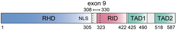
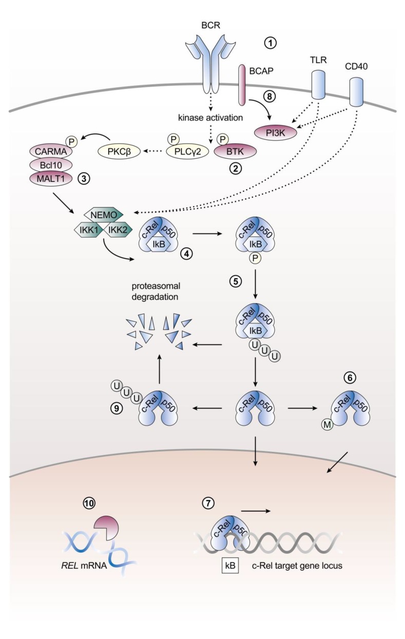
Aberrant constitutive activation of Rel/NF-κB transcription factors is a hallmark of numerous cancers. Of the five Rel family members, c-Rel has the strongest direct links to tumorigenesis. c-Rel is the only member that can malignantly transform lymphoid cells in vitro. Furthermore, c-Rel is implicated in human B cell lymphoma through the frequent occurrence of REL gene locus gains and amplifications. In normal physiology, high c-Rel expression predominates in the hematopoietic lineage and a diverse range of stimuli can trigger enhanced expression and activation of c-Rel. Both expression and activation of c-Rel are tightly regulated on multiple levels, indicating the necessity to keep its functions under control. In this review we meta-analyze and integrate studies reporting gene locus aberrations to provide an overview on the frequency of REL gains in human B cell lymphoma subtypes, namely follicular lymphoma, diffuse large B cell lymphoma, primary mediastinal B cell lymphoma, and classical Hodgkin lymphoma. We also summarize current knowledge on c-Rel expression and protein localization in these human B cell lymphomas and discuss the co-amplification of BCL11A with REL. In addition, we highlight and illustrate key pathways of c-Rel activation and regulation with a specific focus on B cell biology.
Keywords: B cells; DLBCL; FL; NF-κB; PMBCL; REL gene locus amplification; c-Rel; cHL; lymphoma.
Conflict of interest statement
The authors declare no conflict of interest.
Figures
Similar articles
-
The c-Rel transcription factor and B-cell proliferation: a deal with the devil.Oncogene. 2004 Mar 25;23(13):2275-86. doi: 10.1038/sj.onc.1207410. Oncogene. 2004. PMID: 14755244 Review.
-
Expression of TRAF1 and nuclear c-Rel distinguishes primary mediastinal large cell lymphoma from other types of diffuse large B-cell lymphoma.Am J Surg Pathol. 2007 Jan;31(1):106-12. doi: 10.1097/01.pas.0000213334.40358.0e. Am J Surg Pathol. 2007. PMID: 17197926
-
Gains of REL in primary mediastinal B-cell lymphoma coincide with nuclear accumulation of REL protein.Genes Chromosomes Cancer. 2007 Apr;46(4):406-15. doi: 10.1002/gcc.20420. Genes Chromosomes Cancer. 2007. PMID: 17243160
-
c-Rel Is the Pivotal NF-κB Subunit in Germinal Center Diffuse Large B-Cell Lymphoma: A LYSA Study.Front Oncol. 2021 Apr 20;11:638897. doi: 10.3389/fonc.2021.638897. eCollection 2021. Front Oncol. 2021. PMID: 33959502 Free PMC article.
-
Aberrant rel/nfkb genes and activity in human cancer.Oncogene. 1999 Nov 22;18(49):6938-47. doi: 10.1038/sj.onc.1203221. Oncogene. 1999. PMID: 10602468 Review.
Cited by
-
Genetic deregulation of REL in germinal center B cells induces generation of a pool of lymphoma precursor cells.Blood Adv. 2025 Aug 12;9(15):3676-3688. doi: 10.1182/bloodadvances.2023012166. Blood Adv. 2025. PMID: 40267227 Free PMC article.
-
Combination adjuvant improves influenza virus immunity by downregulation of immune homeostasis genes in lymphocytes.Immunohorizons. 2025 Jan 24;9(2):vlae007. doi: 10.1093/immhor/vlae007. Immunohorizons. 2025. PMID: 39849993 Free PMC article.
-
NF-κB Dependent Chemokine Signaling in Pancreatic Cancer.Cancers (Basel). 2019 Sep 26;11(10):1445. doi: 10.3390/cancers11101445. Cancers (Basel). 2019. PMID: 31561620 Free PMC article. Review.
-
Altered pathways and targeted therapy in double hit lymphoma.J Hematol Oncol. 2022 Mar 18;15(1):26. doi: 10.1186/s13045-022-01249-9. J Hematol Oncol. 2022. PMID: 35303910 Free PMC article. Review.
-
Role of B-Cell Lymphoma/Leukemia 11A in Normal and Malignant Hematopoiesis.Biology (Basel). 2025 Jan 1;14(1):26. doi: 10.3390/biology14010026. Biology (Basel). 2025. PMID: 39857257 Free PMC article. Review.
References
-
- Carrasco D., Weih F., Bravo R. Developmental expression of the mouse c-rel proto-oncogene in hematopoietic organs. Development. 1994;120:2991–3004. - PubMed
-
- Harling-McNabb L., Deliyannis G., Jackson D.C., Gerondakis S., Grigoriadis G., Brown L.E. Mice lacking the transcription factor subunit Rel can clear an influenza infection and have functional anti-viral cytotoxic T cells but do not develop an optimal antibody response. Int. Immunol. 1999;11:1431–1439. doi: 10.1093/intimm/11.9.1431. - DOI - PubMed
Publication types
Grants and funding
LinkOut - more resources
Full Text Sources
